Are you having a lump in one of your joints that is paining with activity and causing numbness/tenderness in the associated limb? The problems could be the result of an underlying disorder known as Osteochondroma. Read and know all about the disease, including its causes, symptoms, treatment options, prognosis and more.
Osteochondroma Definition
Page Contents
It is a benign (noncancerous) tumor, comprised of bones and cartilage, which usually arises in children or adolescents. This abnormal lump develops on the surface of a bone located close to the growth plate.
Osteochondroma ICD10 Code
The ICD10 Code for this disease is C40-C41.
Osteochondroma Types
These tumors are classified into two types, depending on their number. Single tumors of this form are referred to as Osteocartilaginous exostosis. More than one tumors of this nature are collectively called Multiple Osteochondromatosis.
Solitary Osteochondroma
These are supposed to be the most common type of benign bone tumor and account for 35-40% of all non-cancerous tumors of the bone. These types of lumps are not cancerous and they do not spread (metastasize) to other regions of the body. In children, a solitary Osteochondroma may arise at the time of growth. This can occur in cases where the bone grows out from the growth plate rather than growing in line with it.
Solitary Osteochondroma tumors are most common at the end of long bones that meet other bone ends to form joints. These kind of lumps are typically found to develop in the shoulder, hip and knees. These types of bony protuberances may have a stem or stalk sticking out from the healthy bone. In case of the presence of a stalk, the structure is referred to as Pedunculated. In cases where the bony projection is attached to the bone having a broader base, the structure is termed Sessile.
Multiple Osteochondroma
As the name indicates, these are more than one Osteochondroma tumors arising on the body of individuals. The location and number of these growths tend to vary. Both sessile and pedunculated types of tumors may arise. In very severe cases, these lumps may lead to an abnormal growth of the bones. Many affected individuals may have knock-knees and ankles, malformed forearms and short stature. These abnormalities help detect severe cases Multiple Osteochondroma in its early stages. Like Solitary Osteochondroma, Multiple Osteochondroma tumors may not be detected until early childhood.
Multiple tumors of this type are more common in males than in females. The risk of a malignant progression of Multiple growths is greater than Solitary growths.
Multiple Osteochondroma tumors are also known by various other names, such as:
- Multiple osteocartilaginous exostosis
- Multiple hereditary exostosis (MHE)
- Familial osteochondromatosis
- Multiple hereditary osteochondromatosis
- Diaphyseal aclasia
Osteochondroma Symptoms
Most individuals with solitary tumors of this type are asymptomatic, meaning they do not exhibit any symptoms. In some cases, however, the symptoms may be manifested long after the development of the tumors. The condition is usually detected in patients aged between 10 and 30 years.
The typical symptoms of solitary forms of this condition include:
Tingling or numbness
These tumors can be situated close to a nerve, such as in a position posterior to the knee. If the tumor exerts pressure over the nerve, sufferers may experience tingling and numbness in the associated limb.
Painless lump
Sufferers typically have a painless lump arising in a region close to the joints, particularly in the shoulder or the knee.
Pain that worsens with activity
These tumors can be situated under a tendon or the tough fibrous tissue that attaches bones to muscles. When these lumps arise in such spots, the tendon may move and even “snap” over the bony tumor – thus resulting in pain for sufferers.
Changes in blood flow
If the tumor exerts pressure over a blood vessel, affected individuals may suffer from changes in the flow of blood. This can result in changes in limb color or loss of pulse. However, it is only in rare cases that these tumors are found to cause changes in blood flow.
In some patients, a trauma (injury) to the stem of a pedunculated Osteochondroma can make it break. This may result in immediate pain and inflammation in the region which the tumor has developed over.
In Multiple Osteochondroma patients, the symptoms are almost similar although more severe in nature than that of Solitary cases. In cases of multiple patients, painless bumps may arise at the region of development of the tumors. If the lumps exert pressure over the nerves, blood vessels or soft tissues, patients may experience pain and a range of other discomforts.
Osteochondroma Causes
It is not known what exactly leads to the development of this tumor. The condition is not believed to have a sexual preference as both males and females are found to be at equal risk of developing it. Also, it has not been found to result from any injury.
Researchers believe a genetic factor to be responsible for the formation of these kinds of growths. The lumps are believed to be related to a gene known as EXT 1. It is not known how a genetic effect is associated with these tumors. Research is still being conducted to determine the actual relationship between Osteochondroma and genetic factors.
Osteochondroma Diagnosis
As most tumors of this type do not result in discomfort or any other symptoms, these are often discovered accidentally when an x-ray is conducted to diagnose some other condition.
In case of symptoms, patients visit physicians by themselves or taken to doctors’ chambers (if they are too young). Diagnosis usually begins with a physical examination and consideration of the medical history of sufferers. Doctors ask patients about their general health condition, lifestyle and the symptoms that they experience to have a better idea about the problem. The medical history of sufferers is also taken into account.
During physical exam, doctors look for tenderness in the affected area and check the range of motion in the painful region.
The tests conducted for a detection of these tumors include:
X-rays
These provide physicians with a clear image of dense structures, such as the bone, and reveal the bony growth associated to Osteochondroma tumors. A plain x-ray is quite useful in revealing the presence of multiple growths of this nature. The findings are usually severer than those obtained in the detection of solitary cases.
CT scans
Physicians may use CT scans to define the tumor further. These scans help provide more detailed images, particularly of soft tissues. Physicians can also get cross-sectional images with the aid of these scans.
MRI scans
These may be used to detect the presence of cartilage on the tumor surface. MRI scans also help reveal a malignant change of Osteochondroma tumors, although rare. The scans show a thick cartilaginous covering over the tumor in adults, in case of a malignant change. In adult sufferers, Osteochondroma tumors should be checked for cancer if the lump becomes painful or enlarged.
Biopsy
It is important for detection of cancer. In this process, a sample of the tissue of the tumor is taken and sent for microscopic examination. A physician may administer a local anesthetic into the region to induce numbness and use a needle to obtain a small sample. Biopsies may also be carried out as small operative procedures.
Orthopedic onology evaluation
In cases where patients are found to manifest symptoms of an Osteochondroma lump becoming cancerous, they need to be assessed by Orthopedic onologists – physicians who specialize in the treatment of bone tumors.
The signs and symptoms of a tumor of this type becoming malignant are:
- Pain at the region of development of the growth
- Thickness of cartilage cap extending over 2 cm
- Development of an Osteochondroma growth after puberty
If cancer is suspected, sufferers require a thorough evaluation that involves CT and MRI scans of the growth. CT scan of the chest may also be ordered by physicians to check whether any disorder has been transmitted to the lungs through the bloodstream.
When Osteochondroma tumors become cancerous, they commonly change into a malignant cancerous condition known as Chondrosarcoma.
Osteochondroma Differential Diagnosis
The differential diagnosis for this disorder includes ensuring the absence of similar conditions, such as:
- Chondroma
- Osteosarcoma
- Ollier Disease
- Chondrosarcoma
- Myositis ossificans
- Chondromyxoid Fibroma
- Singular chondrosarcoma
- Benign Parosteal Osteochondromatous Proliferation
Osteochondroma Treatment
The treatment for the disorder depends on the type of the tumor that an individual is having. The treatment options for Osteochondroma growths include:
Nonsurgical treatment
In the majority of cases of solitary growths of this nature, treatment involves careful observation extending over a period of time. To keep track of any changes in the growth, doctors may wish to use x-rays on a regular basis. This is true for both solitary and multiple cases of the tumors.
Surgical treatment
The indications for surgical removal, for both types of tumors, are:
- Pain
- Presence of a large cartilage lap
- Pressure on the blood vessels and nerves
Complete removal of an Osteochondroma involves performing an operative process known as Excision. In this method, the tumor is removed at the level of the unaffected bone. Defects such as knock-knee or ankles may need surgical help to straighten the bone.
If multiple Osteochondroma growths turn malignant, surgical cure will depend on the stage of progression of the cancer. Generally, surgery is used to remove malignant tumors. Often, chemotherapy and radiation therapy are used along with operation.
Osteochondroma Prognosis
The outcome of the condition usually depends on the health of patients, extent of progression of the disorder and how fast treatment has been opted for. With proper and timely treatment, prognosis is usually excellent. The time for turn to daily activities usually varies on the basis of the size and the location of the growth. Patients experiencing some pain or discomfort after treatment should limit some activity. Physicians provide such sufferers with specific directions to guide them to recovery.
Osteochondroma Prevention
As the exact cause for development of Solitary Osteochondroma is not known, physicians have not yet been able to find out a preventive measure for it.
Osteochondroma Pictures
Take a look at these images and get a visual idea about this disorder.
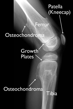
Picture 1 – Osteochondroma
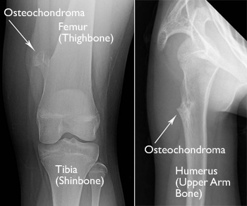
Picture 2 – Osteochondroma Image
If you suspect yourself, or any friend/family member, to have been affected by these tumors get in touch with a medical professional as early as possible. As aforesaid, multiple Osteochondroma growths carry a certain amount of cancerous complication. Hence, it is essential to undergo diagnosis and treatment on an immediate basis to negate such possibilities and elevate the chances of a faster recovery from this discomforting condition.
References:
http://orthoinfo.aaos.org/topic.cfm?topic=A00079
http://www.tumorsurgery.org/patient-education/bone-tumors/bone-tumor-types/osteochondroma.aspx
http://emedicine.medscape.com/article/1256477-overview
http://centerforchildren.med.nyu.edu/osteochondroma
my family suffers from osteochondromas except my dad and grandam