What is Calcific Tendinitis?
Page Contents
It is a form of tendinitis that is characterized by deposits of hydroxyapatite in any tendon of the body. It is generally found in the tendons of the rotator cuff which causes pain and inflammation. This condition could lead to Frozen shoulder.
The calcium phosphate deposits are usually about 1-2 centimeter size. These are usually found in patients who are about 30-40 years old. Diabetic patients develop this more often. The calcium deposits do not always give pain. Even when painful, they usually dissolve within 4 weeks.
Calcific Tendinitis Causes
The prime cause of this disorder is not fully known. Various reasons have been suggested none of which has been conclusively proved. The condition is usually believed to occur due to the deposition of calcium with the rotator cuff tendon but the facts are not clear as to why so happens.
Calcific Tendinitis Stages
This condition usually spreads through some predictable stages and gets resolved without surgery. The stages of progression are as follows:
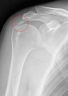
Picture 1 – Calcific Tendinitis
Pre-Calcification Stage
In this stage individuals do not show any symptom. The calcifications develop and experience cellular changes which make the tissues prone to developing calcium deposits.
Stage of Calcification
During this stage, calcium is released from cells and then gets accumulated as calcium deposits. The calcium looks chalky but it is not a solid piece of bone. Once the calcification is completed a resting phase begins which may last for a certain length of time. A resorptive stage, which is the most painful phase of calcific tendonitis, follows this stage. During this process, calcium deposits take the appearance of toothpaste.
Post-Calcification Stage
This is a painless stage in which the calcium deposit goes away and is replaced by a normal looking rotator cuff tendon.
Calcific Tendinitis Symptoms
The pain experienced in this condition is usually unbearable and feels like a constant nagging thing. It is felt in the shoulder, outer upper arm, sometimes down the arm to the hand where it gets maximized due to elevation of the arm. The pain at night gets intensified when the arm is raised. The pain often feels like a tingling sensation in the arm and fingers in certain cases. Other discomforts experienced by sufferers include stiffness of the arm, snapping and weakness of the shoulder.
Calcific Tendinitis Diagnosis
The calcium deposits can be observed with the help of plain X-rays. They appear as discrete lumps or cloudy areas. In X-ray images, they appear cloudy if in the process of re-absorption. This is also the time when they cause the most pain. The deposits are crystalline in nature when they are in their resting phase. Ultrasound tests help in locating the small calcific deposits which can be missed on x-rays. It also helps in estimating the size of the deposits in all directions.
Calcific Tendinitis Treatment
The conservative treatment options for this disorder include:
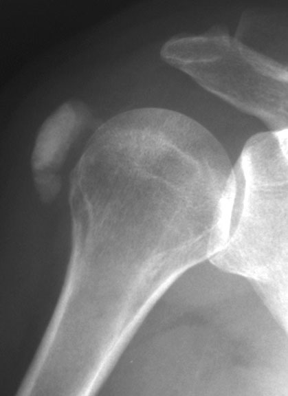
Picture 2 – Calcific Tendinitis Image
Anti-inflammatory Medications
Non-steroidal anti-inflammatory drugs (NSAIDs) are often prescribed to patients with this condition. The analgesic properties of the medicines are helpful in treating this condition.
Physiotherapy
Physiotherapy helps the shoulders to stay strong and keeps irritation at bay. Exercises are prescribed by doctors to maximize the range of motion of the shoulder and enhance muscle strength. Physical therapy modalities, including electroanalgesia, heat and ice therapy, are often carried out to offer relief from the symptoms. These therapies improve the biomechanics of the shoulder and minimize the tension at the affected muscles which allows a decrease in the amount of calcification.
Corticosteroid injections
These injections are administered when the shoulder gets inflamed. It can eliminate pain but does not help in absorbing the calcium. It is helpful in patients as a first treatment measure and is known to yield successful results.
Injections, needle and lavage
Under local anesthesia, the calcium deposits can be broken down by puncturing them continuously with a needle and then dissolving the calcific material with the help of a stream of saline. Most patients receive relief in this procedure.
Extracorporeal Shock Therapy
This therapy makes use of sound waves and focuses on the calcium deposit. It provides relief from pain. In most cases, the deposits disappear or disintegrate.
Surgical Therapy
If the pain does not go with the aid of the above conservative methods, surgery may be opted. An open or an arthroscopic approach may be opted for surgical treatment. Arthroscopic procedure offers a better result. Preoperative ultrasonic localization and probing with a needle have proved successful. Once the deposits are located they can be teased out of the tendon with a hook through a longitudinal cut in the tendon. The Subacromial space is irrigated fully.
In an open surgery, the tendon is cut in a similar fashion and the deposit is drained out. The adjacent tendon edges are trimmed and re-approximated. Post-operation, a sling is used to protect the shoulder from sudden injuries.
This operation has a high success rate in 90 percent of the cases. The rest 10 percent cases might require re-operation. If the deposits are larger in amount, patients are likely to need a repairing of the rotator cuff to fix the defect left in the tendon when the deposit is done away with. The tendon may also be reattached to the bone if the deposit is found to occur at the region where the tendon gets inserted into the bone.
References:
http://emedicine.medscape.com/article/1267908-treatment#a1128
http://en.wikipedia.org/wiki/Calcific_tendinitis
http://orthopedics.about.com/od/rotatorcuff/a/calcific.htm
http://www.shoulderdoc.co.uk/article.asp?section=10
http://www.shoulderinstitute.co.za/calcific_tendinitis.php