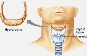
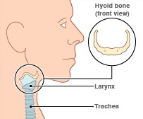
The hyoid bone is a distinct bone that does not join directly to any other human bone. In stead, it suspends in a network of muscles and ligaments.
Hyoid bone Location
Page Contents
The hyoid bone is found in the throat region immediately beneath the person’s chin.
Functions of hyoid bone
The function of this type of bone is to offer support to the tongue, enabling people to appropriately make a number of dissimilar sounds, providing people with oral convenience and tone variation, so individuals can sing at varying intensities.
Original discovery of the hyoid bone
Many and many years back, the hyoid bone was observed in the hominids. A larynx drop, which is a deeper element of the larynx of the gorge, was also seen in the hominids. Larynx drop refers to a development in humans characterized by deep movement of the larynx towards the throat during the process of growing up. Usually, this development occurs during the early years of life. Without the hyoid bone, human being would not be able to communicate suitably as is the care today, virtually making it impossible to decipher what the other people may want to pass across to you. It would be unimaginable if people only made noises and other combinations of long horrible trials. Therefore, it can be safely concluded that the hyoid bone is a very important aspect as far as speech is concerned.
Story behind the discovery of hyoid bone
The original discovery of the contemporary hyoid bone can be traced to Palestine. A Neanderthal guy found in a cave called Kebara was observed to have a unique bone. This made the researchers to develop an interest when they found out that the man had as deeper larynx that had never been witnessed before. They also discovered that the guy had human communication potential. Nevertheless, other discoverers argue that the morphology of this bone is not suggestive of the position of the larynx. It is important to bear in mind the base of the cranium, the mandible as well as the cervical vertebrae besides the skull reference plate.
Origin of the word- Hyoid bone
The word “hyoid” originated from the Greek name hyoeides, which means has the shape of an upsilon. Upsilon is a Greek alphabetic character that considerably resembles the letter U. However, with some leg-like features on the sides the letter also resembles an H having very small legs.
Hyoid bone Structure
The hyoid bone is divided into there main subsections. These are:
- The greater cornu
- The body
- The lesser cornu
The greater cornu of the hyoid bone
Making use of the H to refer to the 3 subdivisions of the hyoid bone can greatly resemble. This can greatly help you to understand the structure of the hyoid bone. The greater cornu is situated on the upper divide of the two perpendicular lines while the less cornu is situated on the lower divide of the two perpendicular lines. On the other hand, the flat line plus the central section between the two perpendicular lines form the body of the hyoid bone.
The body of the hyoid bone
The body of the hyoid bone is made up of four segments.
- The anterior flat is convex-shaped and is stretched frontward and upwards. Well-designed transverse edges having a little downward sloping characterize the upper section of the bone. In a number of scenarios, a perpendicular median edge separates it, thereby making pairs of lateral segments. The section of the perpendicular edge over the slanting line can be observed in most of the samples. However, the lower section can only be observed in uncommon cases. The Geniohyoideus is offered insertion by the anterior plate that can be found in the greater wing of the extended bone segment both below and above the slanting edge, a section of the discovery of the Hyoglossus denotes the lateral circumference of this Geniohyoideus connection. Beneath the slanting edge, there are the key insertions which include Omohyoideus, Sternohyoideus and Mylohyoideus.
- The posterior plate is concave smooth, and stretches downward and backward. The hypothyroid membrane divides it from the nearby epiglottis. In addition, an amount of areola tissues plus a bursa divides it from the nearby hyothyroid membrane.
- The superior margin is circular and provides connection to the adjacent hyothyroid membrane in addition to certain Genioglossus filaments.
- The inferior margin provides medial insertion to the sternohyoideus as well as lateral insertion to the omnohyiodeus. Additionally, it gives an occasional attachment to the thyreohyoideus. There are other muscles that are attached to the hyoid bone and include levator glandulae thyreoideae among others.
During the early stages of development, the lateral margins interact with the superior cornu via synchondroses.
Uniqueness of the hyoid bone
The hyoid bone is distinct because of the way it is situated in the body. While most other human bones will articulate with other bones, the hyoid bone is a clear exception. Instead, it is found suspended in a network of muscles and ligaments. This means that bit creates no links with any bones.
Muscle attachments of the hyoid bone
Some of the superior muscle attachments that link the hyoid bone include:
- Mylohyoid muscle
- Geniohyoid muscle
- Stylohyoid muscle
- Digastric muscle
- Hyoglossus muscle
- Pharyngeal constrictor middle muscle
The inferior muscle attachments of the hyoid bone include:
- Sternohyoid muscle
- Omohyoid muscle
- Thyrohyoid muscle
The hyoid bone is also composed of the greater cornua, which is also referred to as cornua majora or the thyrohyals. The greater cornua extend rearward starting at the lateral margin of the main body of the hyoid bone. Moreover, they are leveled from over downwards and decrease in size as it extends from the rear side. All these projections terminate in a fixed tubercle that is connected to the hyothyroid muscle on the lateral side of the main body.
The upper plate is rough near the lateral margin to provide great potential for attachments of various muscles. These muscles have diverse insertions that make them connect easily to the various surfaces of the hyoid bone. For instance, at the medial margin you’ll find an attachment of the hyothyroid membrane. In addition, the Thyreohyoideus attaches itself to the lateral margin of the bone on the anterior segment.
The lesser cornua are also referred to as the cornua minora or Ceratohyals. They are dual, tiny conical projections connected via their bottoms to suitable angles of joint between the greater cornua and the main body. They are joined to the main body by a network of fibers, and in some cases, they can be connected via discrete diarthrodial junctions. While these joints may be long-lasting, at times they can turn out to be ankylosed.
The lesser cornua can be found in row of the slanting edge on the main body. They seem as morphological extensions. The tip of all the lesser cornu provides a base for attachment of the stylohyoid fiber. Additionally, the Chonroglossus begins at the medial section of the bone’s base.
Ossification of the hyoid bone
Ossification of the hyoid bone starts from 6 locations. These are 2 for the main body while each cornu takes one.
Ossification starts at the section of the greater cornua extending towards the terminal of fetal stage, soon after in the main body.
Ossification in the cornua minora starts at least one year after childbirth but before the start of the third year.
Hyoid bone in animals
Examples of animals that have distinct hyoid bones include the cat species. The four species of the cat i.e. the leopard, jaguar, lion and the tiger are able to produce certain sounds characteristic of deeper larynx in their throats.
Only the aforementioned cat species are able to roar due to development of anatomical structure. The key reason for that can explained this was initially thought to be the partial ossification in the hyoid bones. Nevertheless, new research has indicated that the capacity to make these sounds is because of a number of other morphological aspects, particularly the larynx.
The Uncia uncia is a snow leopard species that is often incorporated in the category of panthera but cannot make the roaring sound. While it had a partial ossification of its hyoid bone, the snow leopard doesn’t have the particular larynx morphology.
In loads of other animals, the hyoid bone has other features including several gills in fish and many corneas in reptiles and amphibians.
Hyoid bone syndrome
Hyoid bone problems leading to tenderness near the greater horn of the hyoid bone.
Hyoid bone Fracture
The hyoid bone is strategically placed such that fractures are very rare. In scenarios of murder, a fracture of the hyoid bone is suspected. It indicates strong cases of strangulation or throttling. Throttling can lead to death because it powerfully reduces the passage of air to the person’s lungs. The neck has a number of points that are vulnerable for powerful throttling and include the carotid arteries.
Strangling is a forceful compression in the neck region that can result in nothingness or even fatal consequences. This is because the act can boost the hypoxic situation in the person’s brain.
Deadly strangling normally takes place in scenarios such as accidents, violence as well as the secondary lethal systems like hanging provided that the neck fails to break. While strangling may not lead to fatal consequences, it is a dangerous situation. Unless interrupted as done in cases of the asphyxias or that of the choking game, strangling can cause early death. However in these events strangling is used in sport and self defensive techniques. Generally, strangling is categorized into three depending on the system applied. These include manual strangulation, ligature strangulation and the lethal system of hanging.
Mortality rate with fracture of hyoid bone
A fracture of the hyoid bone is said to happen in between 17 to 70 percent of the death cases that result from manual strangulation .however, these are uncommonly evidenced in the cases of survivors.
Radiological study of the fracture of hyoid bone
Two scenarios are provided of suspected self strangulation in the manual way in ladies aged between the age of 30 and 35 wherein lateral X-rays showed that there is a common but isolated hyoid bone fracture characteristically affecting the greater cornua of the body. Both cases necessitated the use of X-rays since the victims experienced such symptoms as discomfort during swallowing of food and pain when moving the neck even slightly. The other case reported symptoms such as pain when swallowing food and on talking.
Radiological case of the isolated fracture of the hyoid bone is greatly important to criminal court hearings in situations of unsuccessful manual strangulation. The value of this evidence is greatly increased in cases where the authorities delay their presentations while the exterior evidence of manual strangulation or injury on the neck region may be scant or totally invisible. Care should be taken in cases where the signs and symptoms of strangulation continue for a long period. Consideration is also important when taking the X-rays especially if the pain persists.
Nevertheless, the hyoid bone is not yet well-developed in kids and teenagers and so strangulation cases may be highly regrettable.
Reference:
http://www.ncbi.nlm.nih.gov/pmc/articles/PMC1256534/pdf/janat00047-0243.pdf
http://www.returning-home.net/The%20Hyoid%20Bone%20of%20Separation.pdf


No Responses