Actinic keratosis is a chronic dermatological disorder, which can easily progress to invasive carcinoma of the skin. Read on to know all about this disorder, including its causes, symptoms, diagnosis and treatment.
What is Actinic keratosis?
Page Contents
- 1 What is Actinic keratosis?
- 2 Actinic keratosis Incidence
- 3 Actinic keratosis Symptoms
- 4 Actinic keratosis Types
- 5 Actinic keratosis Causes
- 6 Actinic keratosis Pathophysiology
- 7 Actinic keratosis Diagnosis
- 8 Actinic keratosis Differential Diagnosis
- 9 Actinic keratosis Treatment Options
- 10 Natural treatment for Actinic keratosis
- 11 Actinic keratosis Prognosis
- 12 Actinic keratosis Prevention
- 13 Actinic keratosis (Solar Keratosis) Pictures
This is a precancerous condition of the skin marked by a small lesion due to increased exposure to UV rays from the sun. It is also known by other names like:
- Senile keratosis
- Solar keratosis
Actinic keratosis Incidence
The condition accounts for 50% of the global population, and is usually common in fair-skinned individuals between the ages of 30 and 60 years. Males get more affected than females.
Actinic keratosis Symptoms
In this condition, rough, thick, scaly patches can be observed on sun-exposed areas of skin. The most common sites for such thickened scaly growths are the face, specifically on the lips, cheeks, and bridge of the nose and ears. Other areas include:
- Scalp
- Back of the neck
- Upper chest
- Forearms
- Hands
The condition initially manifests as small crusty spots measuring between 2-6 millimeters in diameter that cannot be easily spotted. However, they can be felt by stroking the skin. These feel similar to rubbing sandpaper on touch. In the course of time, the skin lesions grow bigger in size. These may grow up to 3-10 mm or several centimeters, resembling warts. These dry skin patches usually appear red, dark, light, pink, or a combination of all these, with a white or yellowish scale on top. In some cases, there is no demarcation between the tiny bumps and the surrounding skin.
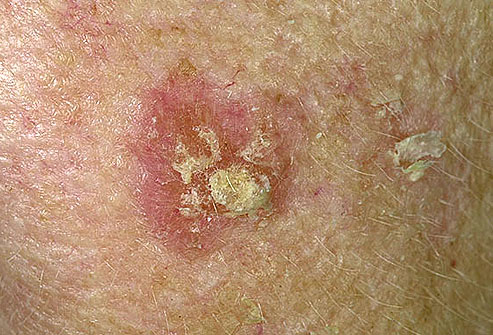
Picture 1 – Actinic Keratosis
The condition may coexist with other manifestations of sun damage like:
- Wrinkles
- Sallowness
- Superficial blood vessels
Many patients experience excruciating pain when the enlarged lesions repeatedly collide with fingers or clothing. The skin becomes tender when multiple lesions develop within a single area, causing more discomfort and agony. Itching and burning in the affected regions are its additional features. A counterpart of solar keratosis, called actinic cheilitis, causes inflammation and cracking of the lips. The lower lip undergoes scaling and becomes rough, with blurring of the line bordering the lip and skin.
Actinic keratosis Types
Following are the specialized forms of Solar keratosis:
Hyperkeratotic actinic keratosis/Exaggerated hyperkeratosis
In this type, the outermost layer of the epidermis consisting of dead cells, becomes hypertrophic and has a rough texture. Development of cutaneous horns, in which the skin has a horn-like projection, is its most prominent feature. The skin protrusions are remarkably thick and the base of the horn could be flat or nodular. The condition typically occurs on the scalp and back of the hands.
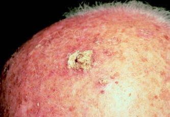
Image 2 – Actinic Keratosis on Scalp
Pigmented actinic keratosis
This is the heavily pigmented variant of the condition often clinically mistaken for lentigo maligna, a flat mottled, brownish malignant growth on the skin due to increased deposition of melanin. Here, the pigmentation occurs in the lower epidermis.
Lichenoid actinic keratosis
This particular version of keratosis is characterized by circumscribed, firm, scaling plaques with rich supply of inflammatory cells beneath the skin. Therefore, the skin papules appear bright red in color.
Atrophic actinic keratosis
Unlike the hyperkeratotic form, the atrophic lesions formed here leads to paper-thin skin, especially in older patients.
Actinic keratosis Causes
The skin lesions are UV light-induced and affect those individuals who get frequently exposed to the sunlight. Sensitivity to UV light is an inherited condition in fair-skinned or blonde patients. Such individuals often complain of sunburns and tans. Prolonged and intense sun exposure acts as a contributing factor in causing the condition. It may take several years or even decades for the skin to undergo keratosis. Short-term exposure to the sun does lead to the premalignant disorder. The epidermis, which is the upper or outer layer of the skin, is made up of flat, scale-like cells. The deeper region of the epidermis comprises of melanocytes, producing melanin for pigmentation of the skin. Normally, old/dead epidermal cells are constantly replaced with healthy cells for proper maintenance of the skin. In this condition, however, the UV rays directly penetrate into the skin and damage it. As the epidermal cells begin to deteriorate, there is a modification in the texture and color of the skin. The condition is more obvious when simple blemishes on the skin transform into painful lesions, having high potential for malignancy. Commercial tanning lamps as well as beds form the potential source for solar damage besides sunlight. The fluorescent lamps used in these indoor tanning equipments radiate UV rays, which could be detrimental to the skin. In addition to long-term sun exposure, some patients are at a greater risk of developing the lesions if they have any of the following conditions:
- Suppressed immune system, due to regular intake of immunosuppressive drugs after an organ transplant
- Psoriasis, treated with PUVA – a therapeutic procedure involving administration of oral drugs called psoralens followed by exposure to UVA light
Key studies have found a correlation between senile keratosis and existence of human papillomavirus in skin. The chronic skin infection, which slowly leads to formation of tumors, has been attributed to the viral agent. In fact, Beta-papillomavirus has been discovered in patients with skin oncogenesis and UV-induced keratosis. However, medical experts are still unaware of the mechanism by which these lethal virus triggers tumor growth.
Actinic keratosis Pathophysiology
Histopathological analysis of solar-damaged skin reveals UV-induced mutations in some genes, including TP53 as well as deletion of the gene encoding p16 tumor suppressor protein that regulate the cell cycle. Under a microscope, the epidermal lesion is filled with atypical, pleomorphic keratinocytes in the basal layer, which extends to the upper granular and cornified layers. The prime characteristics of the epidermal layer include the following:
Acanthosis
It is marked by diffused thickening of the epidermal cells.
Parakeratosis
It is marked by persistence of the nuclei of keratinocytes as they rise into the stratum corneum of the epidermis
Dyskeratosis
It is characterized by abnormal premature keratinization within individual or groups of cells below the stratum corneum.
The degree of cellular change is less pronounced and the lesions are still in the primitive stage. Although the epidermis appears thick, there is a loss of granular layer with wide gaps between the cells. In a few instances, inflammatory cells permeate the epidermal layer and exhibit actinic elastosis, in which accumulation of elastin occurs in the dermis.
Actinic keratosis Diagnosis
History of exposure to the sun is not a valid diagnostic criterion as the condition takes time to develop. However, fair-skinned patients who are into regular outdoor activities or occupations involving extended periods of sun exposure are prone to the disease. Physical observation of the skin cannot help locate the lesion, owing to the minute size of the papules. In such cases, doctors can slightly stroke, scratch or rub the affected area of the skin to palpate these localized lesions. The larger skin bumps are, however, more distinct and easier to identify. Further evaluation of the enlarged thick, scaly growths can be done with a normal skin biopsy. This is performed in order to inspect the current stage of precancerous keratosis and rule out the presence of cancer. During the test, examiners usually detect these lesions on sun-exposed areas of the skin like bare scalp, face and hands. The other dermatologic manifestations such as yellowish skin and wrinkles associated with increased UV exposure can be seen alongside the lesions.
Actinic keratosis Differential Diagnosis
During skin biopsy, Seborrheic keratoses – a benign skin condition marked by greasy, yellow or brown warts, occurring on regions not exposed to sun, needs to be first distinguished from solar keratosis. Similarly, the following conditions are also segregated from the pre-cancerous disorder:
- Basal cell carcinoma
- Squamous cell carcinoma
- Discoid lupus erythematosus
- Non-genital warts
- Porokeratosis
- Bowen’s disease/Bowenoid actinic keratosis
Actinic keratosis Treatment Options
Further progression of the disorder should be stopped, as the lesion is still in the premalignant stage. A variety of treatment approaches are available for remission of lesions. Some of these are:
Cryosurgery
The method involves application of extreme cold in the form of liquid nitrogen, solid carbon dioxide, or argon to destroy the abnormal lesions. Under freezing conditions, ice crystals form inside the tiny lumps and tear them apart.
5-fluorouracil (5-FU)
It is a chemotherapeutic cream that causes reddening and inflammation of the affected skin area prior to falling off of the lesions. Post-application, the skin appears smooth and lesion-free. The treatment procedure is, however, discomforting and the skin may remain in the inflamed state for many weeks. Thus, it must be used only in severe cases.
Ingenol mebutate
It is a common constituent of sap in plant and a powerful inducer of cell death. Affected individuals can use gels containing 0.05% or 0.015% of the substance on trunk, upper extremities, face and scalp. Normally, it takes 2 days for the 0.05% gel to show positive results, whereas the 0.015% gel requires minimum 3 days for complete treatment of the condition.
Diclofenac sodium
This cream is laden with a non-steroidal anti-inflammatory drug, analogous to ibuprofen. The topical medication is usually prescribed for at least 60 to 90 days. Its effect is milder than 5-FU and causes less inflammation.
Imiquimod (Aldara)
It is a topical medication that functions as an immune stimulator and fights against the viral agents.
Photodynamic therapy
The first step of the clinical procedure uses an intravenous drug or dye called aminolevulinic acid for sensitizing the skin to light. After a brief incubation period, the target skin is exposed to light from a laser or other source. The photosensitizing medication gets activated and releases an oxygen molecule, which directly destroy the lesions. After the completion of treatment, patients must avoid exposure to sun or fluorescent light for two days to prevent complications.
Laser therapy
Carbon dioxide or Er:YAG lasers can repair the sun damaged skin in a short span of time.
Electrocautery
In this technique, a metal probe heated by electric current is used to excise the skin lesions.
Various other types of surgery
Sometimes, physicians prefer other means of surgical procedures to remove the painful lesions.
Natural treatment for Actinic keratosis
There are certain herbal remedies like green tea and milk thistle that help in alleviating the painful symptoms of this condition. These herbal formulations also protect the skin from the damaging effects of UV rays.
Actinic keratosis Prognosis
Late diagnosis as well as treatment can transform the condition into carcinoma of the squamous cells that forms the surface of the skin. The cancer is capable of spreading to other regions of the skin. However, this seldom occurs in the case of keratotic lesions unless the tumors occupy the deeper layers. Post-treatment, patients must go for annual check-up to make sure that the problem does not recur or the minute lesions are not growing bigger.
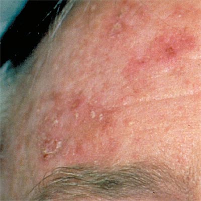
Photo 3 – Actinic Keratosis
Actinic keratosis Prevention
Individuals who are at an increased risk of developing the condition must strictly adhere to the following remedial measures:
- Restrict outdoor activities, especially from 10:00 am to 3:00 pm
- Avoid using artificial tanning beds or lamps
- Apply an appropriate sunscreen or other skincare products on solar damage-prone areas before going out in the sun
- Eat a healthy diet rich in vitamin D and other supplements
- Wear long sleeves, pants, hat/cap and sunglasses to protect against the damaging UV rays
Actinic keratosis has a high rate of malignant transformation and must be immediately diagnosed if the skin spots undergo a sudden change. Healthcare professionals must educate their patients about this condition and instruct them to take the essential measures when out in the sun.
Actinic keratosis (Solar Keratosis) Pictures
Have a look at some of the pictures of Actinic Keratosis for a better understanding.
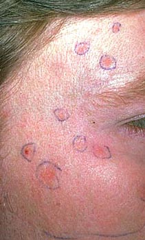
Picture 4 – Actinic Keratosis
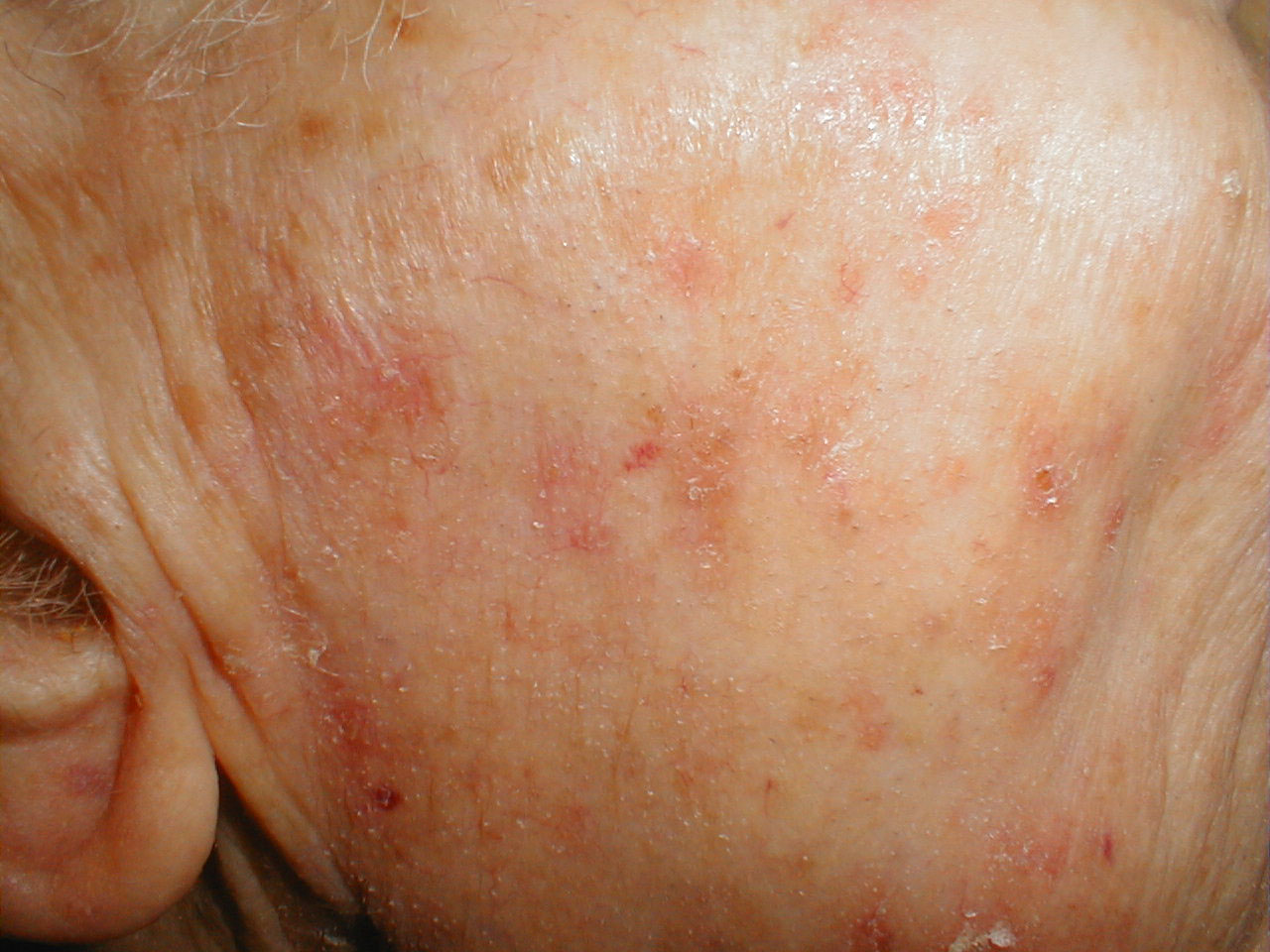
Image 5 – Actinic Keratosis
References:
http://en.wikipedia.org/wiki/
http://www.ncbi.nlm.nih.gov/
http://www.mayoclinic.com/
http://www.skincancer.org/