Angiokeratoma Definition
Page Contents
It is a non-malignant lesion made up of capillaries that gives rise to small deep-red to blue-black marks on the skin. The condition is manifested by hyperkeratosis or thickening of the outermost layer of the epidermis. The vascular lesions could either be single and discrete, or arranged in clusters, and normally do not blanch on being applied with pressure.
Angiokeratoma Types
The benign condition is classified into several types based on the location of these warty elevations. These include:
Sporadic angiokeratoma
In this type, solitary lesions are randomly present on the lower extremities of the body. It is a common dermatological disorder in middle-aged adults.
Fordyce Angiokeratoma
The condition is usually marked by a single lesion or multiple papular growths on the scrotum of males. The small, raised, dark red-to-purple spots may also appear on the shaft of the penis, the inner thigh, or the lower part of the abdomen.
Angiokeratoma circumscriptum
Abnormal clusters of blood vessels that occur during fetal development can unilaterally manifest into a group of lesions on the torso or lower leg, and sometimes on the thighs. As time passes, the lesions may undergo change in shape and size. The condition is rare, but has morphologic similarities to malignant melanoma or pigmented basal cell carcinoma.
Vulvar Angiokeratoma
In this type, solitary or multiple lesions occur on the vulva of females who are above the age of 50.
Angiokeratoma of mibelli
The condition is characterized by 1-5 mm red, vascular papules on the skin. In the later stages, the surface of the lesions thickens to give a scaly or warty skin. This cutaneous, vascular disorder is also known as “Mibelli’s angiokeratoma” or “Telangiectatic warts”.
Angiokeratoma Symptoms
The main clinical presentations of the condition are given below:
Telangiectasias
Small, dilated blood vessels, measuring 0.5-1 mm in diameter, are extensively found near the surface of the skin or mucous membranes. They can develop anywhere on the body, but are commonly seen on the face, particularly on the nose, cheeks or chin.
Acanthosis
In affected patients, the stratum spinosum of the epidermis undergoes thickening, and leads to formation of warts on the skin and mucosal layers of the mouth.
Angiokeratoma Causes
This dermatological condition has often been associated with angiokeratoma corporis diffusum/ Fabry syndrome, a rare, X-linked recessive disorder marked by a deficiency of alpha-galactosidase A. The lysosomal enzyme is needed to metabolize lipids, fat-like substances that include oils, waxes, and fatty acids. In Fabry syndrome, however, mutation in the gene that controls alpha-galactosidase causes accumulation of globotriaosylceramide (Gal-Gal-Glc-Cer) in the vascular endothelium, smooth muscle cells, kidney, myocardium, and nervous system. The symptoms usually arrive before puberty and include:
- Dilation of blood vessels in the genital areas
- Edema
- cardiomegaly
- Diffuse nodularity of the skin
- Casts in the urine
Angiokeratoma Diagnosis
Physical inspection of the affected skin with a dermatoscope may aid in determining the nature of the lesions. For further investigation, biopsy of the abnormal growth can confirm the condition and rule out the presence of other skin disorders. Patients with suspected Fabry’s syndrome may require a simple blood test to measure the level of alpha-galactosidase. A kidney biopsy is normally advisable to assess the amount of intraglomerular lipids.
Angiokeratoma Treatment
The severity of the vascular lesions can be reduced by using the following therapeutic methods:
Surgical excision
The affected tissue is normally removed by using a scalpel prior to skin grafting.
Laser therapy
Carbon dioxide lasers are used to vaporize and seal the affected layers of the skin. This non-invasive technique promotes development of a new layer of skin in the treated area.
Cryotherapy
The method involves the application of extreme cold to destroy the diseased tissues. In this procedure, liquid nitrogen or argon gas flows into a needle-like applicator, which is placed in contact with the damaged skin. In this way, the painful lesions get eradicated to give rise to an improved skin.
Electrocautery
In this procedure, a metal probe heated by electric current is used to destroy the abnormal vascular tissues.
Angiokeratoma is not a fatal condition and may remain asymptomatic for a long time. In most cases, however, treatment becomes necessary due to persistent bleeding or cosmetic concerns.
Pictures of Angiokeratoma
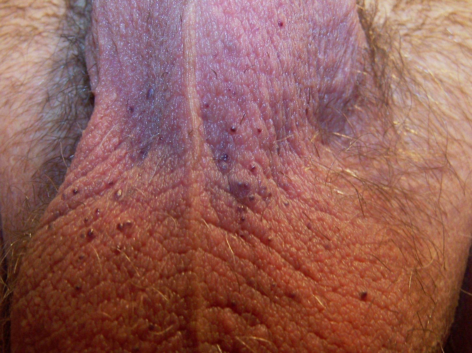
Image 1 – Angiokeratoma of the Scrotum
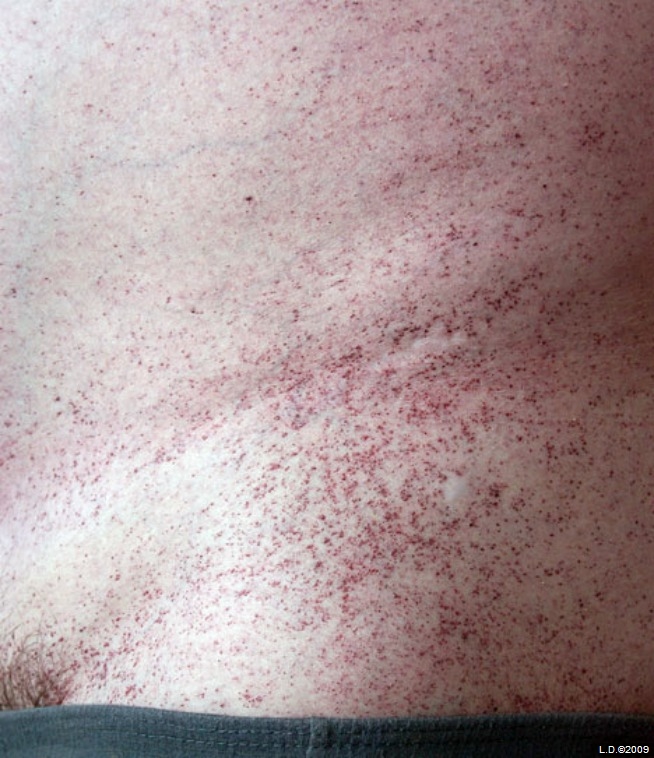
Image 2 – Angiokeratoma
References:
http://emedicine.medscape.com/
http://en.wikipedia.org/wiki/
http://www.onlinedermclinic.
http://dermnetnz.org/vascular/