What is Cerebral Aneurysm?
Page Contents
- 1 What is Cerebral Aneurysm?
- 2 What Happens in Cerebral Aneurysm?
- 3 Cerebral Aneurysm Causes and Risk Factors
- 4 Cerebral Aneurysm Locations
- 5 Cerebral Aneurysm Classification
- 6 Cerebral Aneurysm Symptoms
- 7 Cerebral Aneurysm Diagnosis
- 8 Cerebral Aneurysm Treatment
- 9 Cerebral Aneurysm Complications
- 10 Cerebral Aneurysm Prognosis
- 11 Cerebral Aneurysm Prevention
Cerebral Aneurysm is a cerebrovascular condition characterized by localized dilation of blood vessels. The condition can be either asymptomatic or can cause serious symptoms. In some cases, the disease might even be fatal. The diagnosis of this disease should be followed by immediate medical attention in order to ensure the survival of the patient.
A ruptured cerebral aneurysm, commonly referred to as a subarachnoid hemorrhage, generally requires advanced surgical treatment. Cerebral aneurysm is also known as brain aneurysm or intracranial aneurysm.
What Happens in Cerebral Aneurysm?
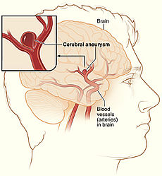
It is a type of cerebrovascular disorder in which the wall of brain artery develops a weak bulge, resulting in the formation of a balloon-like structure on the inner tube of a blood vessel. After a point of time, the flow of blood within the artery rams against the thinned area of the wall, causing wear and tear of the arteries that leads to aneurysms. As the artery walls gradually become thinner due to the dilation, the flow of blood forces the already weakened wall to be swollen. The pressure can cause rupture of the aneurysm, leading to escape of blood in the space around the brain of the patient. This is known as hemorrhagic stroke.
Cerebral Aneurysm Causes and Risk Factors
Aneurysms might occur from congenital defects or from preexisting conditions like atherosclerosis, head trauma or high blood pressure. Research has been conducted to identify the genetics of cerebral aneurysm; various locations have been identified recently which include 1p34-36, 11q25, 7q11, 2p14-15 and 19q13.1-13.3.
Picture 1 – Cerebral Aneurysm
The tendency to develop aneurysms may be inherited. It may also result due to the hardening of arteries and ageing. Some of the risk factors that are responsible for brain aneurysms can be monitored, while others cannot be controlled. The following factors can increase the potentiality of developing aneurysms or rupturing of existing aneurysms.
Family history
Individuals having family history of cerebral aneurysms are highly prone to develop aneurysms compared to those who don’t.
Gender
Compared to men, women are more susceptible to develop brain aneurysms or suffer from a subarachnoid hemorrhage.
Previous case of aneurysm
Patients of a previous brain aneurysm are at the risk of developing aneurysms again.
Hypertension
Individuals having hypertension or high blood pressure are more than likely to suffer subarachnoid hemorrhage.
Race
African Americans have a high probability to have subarachnoid hemorrhage.
Smoking
Smoking greatly increases the risk of cerebral aneurysm rupture.
Other factors that might contribute to the formation of cerebral aneurysms include certain conditions, such as:
- Polycystic Kidney Disease
- Ehlers-Danlos Syndrome
- Fibromuscular Dysplasia
- Marfan Syndrome
- · Polycystic kidney disease
- · Abnormally narrow aorta
- Arteriovenous malformation (AVM)
- Tumors
- Traumatic head injury
- Infections
- Use of drugs, like cocaine
Cerebral Aneurysm Locations
One of the most common locations for these aneurysms to develop includes the arteries located at the basal area of the brain, in an area known as Circle of Willis. Nearly 85% of brain aneurysms form in the anterior portion of Circle of Willis, involving the internal carotid arteries as well as the major branches which supply the middle and anterior sections of brain. Most usual sites include:
- Anterior communicating artery
- Anterior cerebral artery
- Bifurcation of the posterior communicating artery and internal carotid
- Bifurcation of basilar artery
- Bifurcation of middle cerebral artery
- The remaining of the posterior circulation arteries
Cerebral Aneurysm Classification
These aneurysms are classified according to their sizes and shapes. The smaller and medium types of aneurysms are less than 15mm in diameter. The larger ones are classified into large aneurysms (15mm to 25mm), giant aneurysms (25mm to 50mm) and super giant aneurysms, which are more than 50mm in diameter.
The Saccular aneurysms are aneurysms that have a saccular outpouching; they are least occurring form of brain aneurysms.
The Berry aneurysms are also saccular aneurysms but their stems or necks resemble a berry.
The Fusiform aneurysms are the ones without stems.
The Hunt and Hess scale grades the symptoms of ruptured brain aneurysms according to the severity of subarachnoid hemorrhage.
- Grade 0: These are incidentally discovered, unruptured and asymptomatic aneurysms.
- Grade 1: This includes mostly asymptomatic aneurysms, which can sometimes cause minimal headache and a slight nuchal rigidity.
- Grade 2: Aneurysms of this grade can cause moderate to intense headache and nuchal rigidity with no neurological deficiency except the 6th nerve palsy.
- Grade 3: Aneurysms of the grade 3 are drowsy, causing minimal neurologic deficit.
- Grade 4: These cerebral aneurysms are stuporous, causing moderate to intense hemiparesis; early decerebrate rigidity along with vegetative disturbances can be seen.
- Grade 5: These include the most severe cases of aneurysms that can cause deep coma and decerebrate rigidity. They can also be fatal.
According to the appearance of the subarachnoid hemorrhages, the Fisher Grade classifies aneurysms into:
- Grade 1: There is no presence of hemorrhage.
- Grade 2: The subarachnoid hemorrhage is less than 1mm thick.
- Grade 3: The subarachnoid hemorrhage is more than 1mm thick and has the highest probability of vasospasm.
- Grade 4: This includes subarachnoid hemorrhage of any thickness accompanied by intra-ventricular hemorrhage or parenchymal extension.
Unlike Hunt and Hess scale, the Fisher scale is not meant to be a prognostic scale, and is more useful in illustrating the occurrence of subarachnoid hemorrhage and the risks for vasospasm.
Cerebral Aneurysm Symptoms
An individual having an aneurysm may not have any symptoms. Especially an unruptured cerebral aneurysm may go totally asymptomatic. On the other hand, intense headache may be experienced by a patient if the aneurysm starts leaking blood. A sentinel headache occurs as a sign of an oncoming rupture. Symptoms may arise if an aneurysm pushes on the nearby structures inside the brain or ruptures, thereby causing bleeding in the brain. Symptoms also depend on the location of the aneurysm, whether or not it ruptures and which part of the brain it is actually pushing.
The various symptoms of cerebral aneurysm include the following:
- Eye pain
- Stiff neck
- Neck pain
- Headaches
- Loss of vision
- Double vision
A sudden and severe headache is an indication of a ruptured aneurysm. The other symptoms of a rupture include:
- Seizures
- Lethargy
- Dizziness
- Confusion
- Loss of vision
- Double vision
- Dilated pupils
- Drooping of eyelids
- Speech impairment
- Sleepiness or stupor
- Occasional stiff necks
- Loss of consciousness
- Certain infections of blood
- Heavy consumption of alcohol
- Photophobia or sensitivity to light
- Numbness in some parts of the body
- Lowered levels of estrogen after menopause
- Headaches accompanied by vomiting and nausea
- Sudden changes in mental conditions or awareness
- Muscle weakness and difficulty in moving various parts of the body
Cerebral Aneurysm Diagnosis
As an unruptured cerebral aneurysm frequently does not give rise to any symptoms, patients are often diagnosed with the condition while undergoing treatment for a different health disease. If a doctor suspects a case of brain aneurysm, he might recommend the following diagnostic tests:
- Computed Tomography Scan or CT scan
- Computed Tomography Angiogram Scan or CTA scan
- Magnetic Resonance Angiography or MRA
- Cerebral Angiogram
- Cerebrospinal fluid test
- Electroencephalogram or EEG
Cerebral Aneurysm Treatment
An emergency treatment for patients having a ruptured brain aneurysm normally includes restoring the deteriorating respiration as well as reducing intracranial pressure. At present, there are two methods for treating intracranial aneurysms:
- Surgical clipping
- Endovascular coiling
Either of these two procedures is executed within 24 hours after the onset of bleeding to pause rupturing of the aneurysm and minimize the risk of bleeding again.
The doctor normally considers several factors while deciding on the optimum treatment option for a patient. These normally include:
- The age of the patient
- The size of an aneurysm
- Additional risk factors, if any
- The overall health condition of the patient
As the potential of rupture is low in case of a small aneurysm, and surgery for cerebral aneurysms is always risky, a doctor might want to observe the condition for some time instead of immediately opting for surgery. However, if a patient already has a history of ruptured aneurysm or if the aneurysm is large in size, a physician would recommend surgery immediately.
The two methods for cerebral aneurysm repair are described below:
Surgical Clipping
This procedure was introduced in 1937 by Dr. Walter Dandy from Johns Hopkins Hospital. It involves the performance of a craniotomy and exposing the aneurysm, after which a small metallic clip is placed around the base of the aneurysm in order to separate it from the normal blood circulation. This reduces the pressure and prevents the aneurysm from rupturing. Surgical clipping of cerebral artery aneurysm shows lower rate of recurrence of aneurysm after treatment.
Endovascular coiling or Coil embolization
This treatment procedure was introduced in 1991 by Guido Guglielmi at the UCLA. It involves passing a thin catheter into the femoral artery of the groin, through aorta into the cerebral arteries and then finally into the aneurysm itself. Once the catheter reaches the aneurysm, a number of platinum coils are released into the aneurysm. These coils kick off a thrombotic or a clotting reaction inside the aneurysm that can prevent any further bleeding from the particular aneurysm. A small incision is made through which the catheter is inserted. While dealing with broad-based aneurysms, the doctor may insert a stent inside the parent artery which may then serve as scaffold for the coiling. This process is known as stent-assisted coiling.
In some occasions, bulging of certain aneurysms make it necessary for the doctor to cut out the aneurysm and stitch together the endings of the blood vessels. However, such cases are very rare. Also, an artery may be not long enough to be stitched together. In such cases, another artery needs to be used.
A doctor may also recommend several medications and other forms of treatment to manage the condition. These normally include:
- Pain relievers, like acetaminophen, for relieving headache
- Calcium channel blockers like nimodipine to stop calcium from entering the cells of blood vessel walls and reduce vasospasm
- Intravenous injections of vasopressors
- Anti-seizure medications like levetiracetam and phenytoin
- Angioplasty for prevention of strokes
- Lumbar or ventricular draining catheters along with shunt surgery to reduce cerebral pressure caused by excess cerebrospinal fluid
- Rehabilitative therapies, for helping the patient cope with the challenges posed by the associated symptoms
Cerebral aneurysms which cause bleeding can be very serious. In many cases, these ultimately lead to disability or death. Management of the condition requires hospitalization, accompanied by intensive care for relieving cerebral pressure, avoiding re-bleeding and maintenance of vital functions such as breathing and blood pressure.
Cerebral Aneurysm Complications
The following complications might result from cerebral aneurysm:
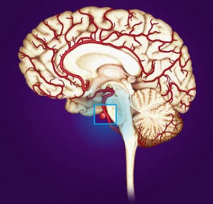
Picture 2 – Cerebral Aneurysm Image
- Stroke
- Seizures
- Deep coma
- Re-bleeding
- Hyponatremia
- Hydrocephalus
- Multiple aneurysms
- Cerebral Vasospasm
- Loss of consciousness
- Intracranial hematoma
- Subarachnoid hemorrhage
- Rupture and bleeding of aneurysm
- Increase in pressure within the skull
- Numbness of different parts of the body
Cerebral Aneurysm Prognosis
The prognosis of such an aneurysm depends on its location and extent, patient’s age, neurological conditions and general health. Some individuals having a ruptured brain aneurysm die from initial bleeding. Others may recover with only little or absolutely no neurological deficiencies. Generally, patients of Hunt and Hess grade 1 and 2 hemorrhage and younger patients might expect a good prognosis without permanent disability or death. Older patients as well as those with poorer grades in Hunt and Hess scale have poorer outcomes. Around 10% of patients having a cerebral aneurysm rupture expire before even receiving medical care. If left untreated, 50% patients are going to die within 1 month. Early diagnosis, followed by appropriate treatment is necessary to guarantee a better prognosis.
Cerebral Aneurysm Prevention
There are no known ways by which the formation of berry aneurysms can be prevented. Treating a patient for high blood pressure might reduce the chances of rupturing of an existing aneurysm. Controlling atherosclerosis risk factors might reduce the possibility of some forms of aneurysms. If discovered early, a non-ruptured aneurysm can be managed before it can cause any problems.
References:
http://en.wikipedia.org/wiki/Cerebral_aneurysm
http://www.webmd.com/brain/tc/brain-aneurysm-topic-overview
http://www.ninds.nih.gov/disorders/cerebral_aneurysm/cerebral_aneurysms.htm
http://www.mayoclinic.com/health/brain-aneurysm/DS00582
http://emedicine.medscape.com/article/1161518-overview


