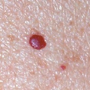
Cherry Angioma is a common skin condition that affects individuals over 30 years of age. Read on to know what is Cherry Angioma as well as its causes, symptoms, diagnosis and treatment.
What is Cherry Angioma?
Page Contents
- 1 What is Cherry Angioma?
- 2 Polyploid Angioma
- 3 Cherry Angioma Symptoms
- 4 Causes of Cherry Angioma
- 5 Cherry Angioma Diagnosis
- 6 Cherry Angioma Treatment
- 7 Cherry Angioma and Bleeding
- 8 Cherry Angioma Home Treatment
- 9 Cherry Angioma Prognosis
- 10 Cherry Angioma Complications
- 11 Cherry Angioma Prevention
- 12 Cherry Angioma Pictures
It is a benign (noncancerous) growth on the skin surface that comprises of blood vessels. Many people suffer from these growths at a later stage of their life, with the onset generally occurring at an age of over 40. However, younger people can also get these growths.
The disease is also known as “Senile angioma”. The lesions formed in this condition are also referred to as “Campbell de Morgan spots”, named after the noted British surgeon Campbell de Morgan of 19thcentury. Campbell was the first person to note and describe these abnormal skin growths.
Polyploid Angioma
In some individuals, more than one angioma lesions may be found to arise next to each other. Multiple angioma growths adjoining each other are collectively known as “Polyploid Angioma”. These mainly arise on the torso (trunk) and appear as cherry red papules. The growths are seen to develop on the same region and have a tendency to bleed easily due to the many blood vessels that they contain. Even a minor injury to the papules may make them vulnerable to a rupture, resulting in release of blood from them.
Cherry Angioma Symptoms
The condition is characterized by the development of lesions or growths on the skin surface. These are small in size measuring about 1/4 inch in diameter. These are smooth and bright, cherry-red in appearance. The lesions may protrude (stick out) from the skin.
Each of these lesions comprise of several weakened and enlarged blood vessels, encircled by lymph. Often, the growth assumes a dome-like form over the skin with the top being somewhat flattened. The color typically ranges from vibrant cherry red to dark purple. The growths commonly arise on the trunk of the body, particularly on the back. However, they may also originate in other areas of the body. These are quite common skin growths that are found to vary in size.
Causes of Cherry Angioma
Medical researchers are still in the dark regarding the exact cause of formation of Cherry angiomas. They have, however, laid down some possible causes for Cherry angioma development. These are:
Aging
The lesions seem to be associated with increase in age as they are found to arise more in older adults, especially those over age 30.
Chemical Exposure
In some cases, these growths are found to arise in response to an exposure to chemicals, such as 2-butoxyethanol, mustard gas, cyclosporine and bromides.
Heredity
Genetic factors are also held to be responsible for the growth of these lesions as they are found to arise in some cases in members of the same family.
Cherry angiomas are regarded as the most prevalent form of angiomas. Despite this fact, there is a lack of proper knowledge about the causes and possible sources of these growths. This may be attributed to half-hearted attempts by most researchers in the medical field. It is partly due to the fact that these lesions are benign and do not result in major complications.
Cherry Angioma Diagnosis
The diagnosis of this skin condition is mainly based on the physical appearance of the growths. A physical observation of the lesions is usually enough for doctors to detect this syndrome. In most cases, additional tests are deemed to be unnecessary. However, if doctors suspect a more complicated underlying problem, a skin biopsy may be performed to confirm the diagnosis.
Cherry Angioma Treatment
Typically, no treatment for Cherry angioma is necessary. Medical removal is required only in rare cases. Ablation is generally performed by methods like:
- Freezing (Cryotherapy) – The technique involves freezing the lesions by spraying (or applying with a cotton swab) liquid nitrogen over them.
- Burning (Electrosurgery or Cauterization) – It includes inserting an electrical needle into the growths that destroys the blood vessels contained within them. The process may cause scarring.
- Shave excision – The process allows lesions to be removed delicately with a blade and also helps in histological confirmation of diagnosis.
- Pulsed Dye Laser (PDL) therapy – The method uses an intense beam of concentrated light to destroy blood vessels within the growths. Following therapy, the lesions develop into crusts and fall of naturally from their original region of formation.
Traditionally, Electrosurgery or Cryosurgery has been used for removing these growths. These are benign growths that pose no risks to sufferers. However, some patients may prefer Cherry angioma removal due to cosmetic reasons. The growths are cosmetically unattractive, especially when they appear on face. In some cases, these are also found to bleed frequently. Some patients may also suffer from discomforts if the lesions arise in some region that creates problems while carrying out daily activities. Growths in uncomfortable spots may also be at risk from getting pressed by things like straps of backpacks or bags, bra straps and waist bands. It may be a good idea to remove the lesions in such cases and make the patient feel more comfortable.
Cherry Angioma and Bleeding
Cherry angiomas comprise of a large number of blood vessels. Due to this, they tend to release blood freely on getting injured. Naturally, it is not advisable to try and puncture a lesion of this type. Bleeding may also occur if the growths are subjected to pressure or stress, as is probable if the bump gets entrapped within the folds of clothing. If patients notice bleeding from their Cherry angioma lesions, they should use warm soapy water to wash the area gently. Slight pressure may be applied with a cotton pad or ball until bleeding reduces. The area may be lightly wrapped with a bandage to prevent seepage of blood onto clothing.
How to Remove Cherry Angioma with Laser Therapy?
Intense Pulsed Light (IPL) or pulsed dye laser has recently been put to use for the treatment of these skin growths. The treatment is usually permanent and helps remove the lesions permanently. However, new growths tend to arise over time in new locations though they may require several years or more to grow large enough to be noticeable. These lesions may also be removed with laser therapy. Laser removal is a speedier process and helps remove many spots within a very short span of time.
Cherry Angioma Home Treatment
At home, Cherry Angioma removal can be done with the aid of natural methods like:
Sandalwood-basil paste
Mix sandalwood powder with fresh basil leaves and make them into a paste. Apply this on the skin. This will help keep the pores clean and free from getting blocked by oils from skin. Sandalwood cleanses and moisturizes skin and keeps it naturally free from germs.
Turmeric powder paste
Mix 5 tablespoons of turmeric powder with equal amount of sandalwood powder and enough rosewater. Form the mixture into a paste and apply over the lesions. Wash this off after half-an-hour.
Water
Drink a minimum of eight glasses of water every day. This will flush toxins out of the body, which may get trapped under the skin and obstruct pores.
Vitamin supplements
Intake of supplements of Vitamins A and E helps improve overall health and maintain healthy skin.
Witch Hazel
Bacteria are a major agent that obstructs skin oil and clogs skin pores. Witch hazel reduces bacteria and cleans the skin.
Dietary modifications
Including more fruits and vegetables and avoiding junk foods can help cleanse the system and remove toxins from the body. This can have a positive effect on the skin.
Natural treatment usually takes time to show effects. Unlike artificial treatment, however, they produce no side effects and cure the growths permanently.
Cherry Angioma Prognosis
These are noncancerous (benign) and usually harmless, except for very rare cases that are characterized by appearance of multiple lesions which may be indicative of a progressing internal malignancy. Generally, removal of the lesions does not result in scarring. In majority of sufferers, the size and number of these lesions increase with advancing age.
Cherry Angioma Complications
The condition is benign and therefore, does not cause any complications. Some of its possible negative developments can be:
- Bleeding, in case of an injury
- Psychological distress
- Changes in appearance
Angiomas are not regarded to be harmful by themselves. However, they can arise as a consequence of other underlying disorders such as cirrhosis or liver disorder. Due to this reason, they should not be treated seriously and never ignored.
Cherry Angioma Prevention
There is no known way to prevent angiomas. Stress is regarded as a major causative factor for the presence and growth of these lesions. Controlling stress with the help of yoga, meditation or other techniques may help prevent the development of Cherry angiomas.
Cherry Angioma Pictures
Take a look at some Cherry Angioma photos to know how these lesions look like. You will find these cherry angioma images quite useful for reference.
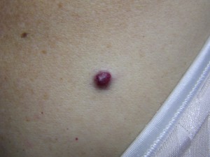
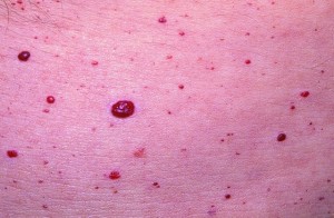
Picture 2 – Cherry Angioma Image Picture 3 – Cherry Angioma Photo
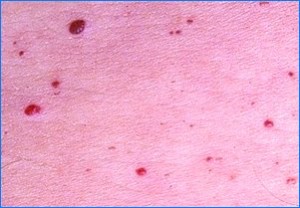
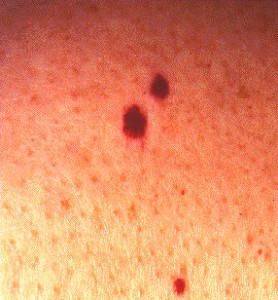
Picture 4 – Image of Cherry Angioma Picture 5 – Cherry Angioma
If you are having Cherry Angioma lesions and notice a change in their appearance or want to have them removed, get in touch with your health care provider immediately. This will help you get treated faster and recover within a few days.
References:
http://www.ncbi.nlm.nih.gov/pubmedhealth/PMH0002413/
http://www.healthscout.com/ency/68/18/main.html

