Cricoid Cartilage is a vital structure of the human body that performs many important functions. Read on to know all about the shape, anatomy, location and functions of this cartilage.
Cricoid Cartilage Definition
Page Contents
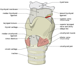
This is a cartilage, or firm elastic tissue that forms the bottom part of the larynx which is more commonly referred to as the voice box. This cartilage is a main element of the structure of the larynx and it provides attachment functions to the ligaments and muscles that co-ordinate function of the Glottis.

Picture 1 – Cricoid Cartilage
Source – wikimedia
Simply put, it is the tiny thick cartilage that forms the posterior and lower sections of the Laryngeal Wall.
Cricoid Cartilage Shape
This cartilage tissue is structurally similar to a signet ring. It is also referred to as Adam’s apple. It is also known simply as “Cricoid”. The word “Cricoid” comes from the Greek word “krikoeides” that means “ring-shaped”. The name is obviously a reference to the ring-like structure of the cartilage. It is the only complete cartilaginous ring that can be found around the trachea.
Cricoid Cartilage Composition
It is composed of Hyaline, another type of cartilage. This is the reason why it can become ossified or calcified, especially when an individual becomes older.
Cricoid Cartilage Location
It is located in the throat. It is the biggest cartilage that can be found in the larynx. It is situated in a position that is just inferior to the thyroid cartilage located in the neck. The median Cricothyroid ligament joins it to the medial position of the thyroid cartilage. The cricothyroid joints attach the Cricoid Cartilage posteo-laterally to this cartilage.
Cricoid Cartilage Vertebral Level
This cartilage is situated in the sixth level of the cervical vertebra in humans.
Cricoid Cartilage Surface Anatomy
The cartilaginous rings surrounding the trachea are inferior to this cartilage. These ring-shaped cartilages are non-constant. They are shaped like the English alphabet “C” with a space in their posterior section. The cricotracheal ligament joins the cricoid to the first tracheal ring. This can be felt between the tough Cricoid and Thyroid cartilages.
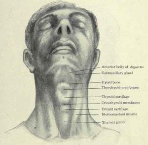
This cartilaginous tissue is anatomically associated with the Thyroid gland. The thyroid isthmus is placed at an inferior position to this tissue. The two lobes of the thyroid expand in a superior position on either side of the cricoids. The expansion continues as far as the thyroid cartilage rests above it.
The posterior section of the Cricoid is a little broader than the lateral and anterior sections. This posterior part is known as the lamina whereas the anterior section is known as the band. This may be a reason why comparison is commonly made between the Signet and Cricoid ring.
Cricoid Cartilage Function
The role of this cartilage is to join various muscles, ligaments and cartilages that are involved in closing and opening of the airways. It is also involved in production of speech. It is to be remembered that cartilage is a very firm connective tissue. It forms a covering over the ends at the joints of bones. It functions as a surface for articulation and allows smooth movement of the joints. Cartilage is not as tough as bone. It is one of the structures of the body that is fully or partially flexible.
When a person suffers from severe respiratory problems and all other attempts fail to bring about any improvement in the condition, a hollow needle may be inserted in the cartilage to assist in breathing. This process is known as Cricothyrotomy or Emergency Airway Puncture.
Cricoid Cartilage Clinical Significance
An Anesthesiologist usually presses on the Cricoid Cartilage when a tube or Cannula is introduced before surgery in a patient under general anesthesia. This squeezes the Esophagus lying behind the cartilage and prevents the occurrence of gastric reflux. This is known in medical terminology as Sellick Manoeuvre.
The Sellick Manoeuvre was regarded as the measure of care for many years throughout Rapid Sequence Induction. The American Heart Association still recommends applying pressure on the cricoid while providing resuscitation by using a BVM and also while carrying out Emergent Oral Endotracheal Intubation. Recent research, however, increasingly indicates that pressure on the Cricoid region may not give as much advantage as was thought earlier.
It is also quite possible that Cricoid pressure may be frequently be applied in an incorrect way. Wrongly applying pressure on the Cricoid area may often move the esophagus laterally, rather than correctly compressing it as is the aim of the Sellick Manoeuvre. A number of studies have demonstrated that incorrect Cricoid pressure can cause compression of the glottis (the vocal apparatus of the larynx) to some degree resulting in rise in peak pressures and lowering of tidal volume. It is due to these recent findings that the once widespread use of cricoid pressure during every rapid sequence intubation is rapidly becoming obsolete.
In some cases, a medical process known as Cricoidectomy may be carried out. This involves partial or complete removal of the Cricoid cartilage in the throat. This is frequently performed to provide relief from obstruction inside the trachea. This usually happens when the Hyalin cartilage located within the Cricoid gets hard and calcified with increasing age and blocks the trachea (windpipe). It may also give rise to pain in individuals.
A CT Scan is usually enough to detect Cricoid Cartilage abnormalities and fractures in the human body. Early diagnosis of problems in this cartilage can help in early cure and a faster recovery.
Cricoid Cartilage Pictures
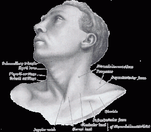
Here are some useful Cricoid Cartilage photos that will give you an idea about the physical appearance of this cartilage within the human body. Take a look at these Cricoid Cartilage images for reference.

Picture 2 – Cricoid Cartilage Image
Source – chestofbooks

Picture 3 – Cricoid Cartilage Photo
Source – wikimedia
References:
http://www.wrongdiagnosis.com/medical/cricoid_cartilage.htm
http://vocalwisdom.com/vocal-physiology-for-singing/larynx/cricoid-cartilage
http://www.viquestwellness.com/ADAM/HIE%20MultiMedia/2/8997.htm
http://www.wisegeek.com/what-is-the-cricoid-cartilage.htm
