A congenital neurological defect, Neural tube defect (NTD) is a complicated condition that gives rise to multiple problems like structural malformations and paralysis. Read and know more about the types, symptoms, causes, diagnosis, treatment and prevention of this debilitating condition.
Neural tube defects Definition
Page Contents
NTD constitutes an array of serious medical conditions of the fetal nervous system that generally occur in its early stages of development in the uterus. It is one of the most common congenital defects of the brain and spinal cord. In the mammalian embryo, neural tube is a tubular structure that eventually gives rise to the central nervous system. In NTD sufferers, the tube does not close completely and leads to an opening in the spinal cord and brain. This leads to serious disability and sometimes, even death.
Neural tube defects Types
The condition can be primarily classified into two forms, which are further categorized into several types.
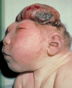
Picture 1 – Neural tube defect
Open NTD
In this form, the entire central nervous system is affected. The neural tissue, covering the brain and spinal cord, is exposed through a defect in the skull or backbone.
It can be further classified into the following types:
Anencephaly
It is the most common form of open NTD where the upper region of the neural tube is not able to close properly. It normally occurs during the 23rd and 26th days of pregnancy, causing loss of a significant portion of the brain and skull. Neonates are born with an absence of a major section of the forebrain – the anterior part of the brain, leading to permanent unconsciousness. In most of these cases, infants are either stillborn or die within a very short span of time.
Encephaloceles
In this form, a gap in the skull leads to sac-like protrusions of the brain and the membranes covering it. An indentation may occur in the mid-section or the back portion of the skull as well as between the nose and forehead. The condition is always associated with other neurological disorders.
Hydranencephaly
In this type, cerebral hemispheres are absent and get replaced by sacs filled with cerebrospinal fluid. Some infants may experience twitching or spasms of the muscles, noticeable at birth.
Iniencephaly
It is rare type of NTD that causes extreme bending of the head to the spine. In affected infants, the neck is generally absent and the face is in the upward direction. The facial skin directly attaches to the chest and the scalp connects to the upper back. There are minimal chances of survival for such newborns. They may die within a few hours after birth.
Spina bifida
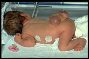
In this developmental congenital disorder some vertebrae overlying the spinal cord are not completely formed and remain open. This condition is additionally classified into spina bifida cystic and spina bifida occulta. In the former condition, the spinal cord develops normally but the meninges that act as a covering of the brain and vertebral column, protrudes from a spinal opening. This is commonly known as “meningocele”. In myelomeningocele, a sac containing a section of the spinal cord and its meninges extrude through the spinal opening.
In spina bifida occulta, a bony defect in the vertebral column causes a cleft that remains covered by skin and may often go undetected during diagnosis. These abnormalities occur when sections of the spinal bones, known as the neural arch and spinous process, are generally non-fatal. It accounts for 10-20% of the healthy population and is asymptomatic.
Closed
It is a rare type of NTD that is normally confined to the spine or vertebral column and does not affect the brain. In this condition, neural tissues are not exposed and a layer of skin covers the spinal defect.
The subgroups of this form include:
Lipomyelomeningocele
It is a form of myelomeningocele with an overlying lipoma- a benign tumor that is composed of adipose tissue.
Lipomeningocele
In this type of meningocele, the spinal cord and lipoma remain within the spinal canal. Neurological defects are visible during the second year of life.
Tethered spinal cord syndrome
In this sub-form, the movement of the spinal cord is restricted within the spinal column due to the abnormal attachment of tissues around the spine at the base. These attachments gradually cause stretching of the spinal cord.
Neural tube defects Statistics
In U.S, NTD is known to affect approximately 1 out of 1000 live births. In Canada, around 260 neonates are born with this condition. Globally, 4000 to 5000 infants are diagnosed with this condition every year.
Neural tube defects Symptoms
The symptoms of this disease may vary from one patient to another. They are also dependent on the type of the disorder that a person is suffering from.
Some of the signs of NTD are:
- Paralysis of the legs
- Partial or total lower body weakness
- Learning disabilities
- Visual impairment
- Hearing impairment
- Stunted growth
- Mental retardation
- Difficulty in movement
- Increased muscle tone
- Respiratory issues
- Progressive enlargement of the head
- Vomiting
- Nausea
- Coma
- Urinary and bowel dysfunction
- Cardiac abnormalities
- Facial defects
Neural tube defects Causes
Health experts are still unaware of the exact cause that disrupts complete closure of the neural tube, resulting in severe physical deformities. However, assumptions of about certain major contributory factors to this condition have been made. These involve:
- Low intake Of folic acid, such as prenatal vitamins
- Ingestion of folate antimelabolites, such as methtrexate (known to impair the function of Folic acid)
- Gestational diabetes in the third trimester of pregnancy
- Obesity during pregnancy
- Presence of Mycotoxins in a wide variety of crops
- Arsenic poisoning from contaminated water
- Elevated body temperature, due to failed thermoregulation
- Excessive exposure to radiation
- Cigarette smoking or exposure to secondhand smoke during maternity
Neural tube defects Diagnosis
Several forms of NTD can be detected during pregnancy with the help of the following tests:
Ultrasound examination
It is generally performed after 18 weeks of pregnancy. In this test, use of high resolution ultrasound may detect any defect in the neural tube of the fetus. Serious types of NTD, such as anencephaly, could be detected earlier than 16 weeks.
Maternal serum alpha-fetoprotein (MSAFP) test
Alpha-fetoprotein (AFP), a plasma protein produced by the fetus, may appear in the bloodstream of a mother after crossing the placenta. High levels of AFP are observed in the case of an Open NTD.
Amniotic fluid alpha-fetoprotein (AFAFP) test
AFP can also be found in the amniotic fluid surrounding the fetus in the amniotic sac. Elevated levels of the protein indicate malformation in the neural tube.
Amniotic fluid acetylcholinesterase (AFAChE) test
Acetylcholinesterase (AChE) is an essential enzyme that maintains proper transmission of impulses between nerve cells and muscles. Prenatal diagnosis of NTD can be performed using amniocentesis or amniotic fluid test. Low levels of AChE in the amniotic fluid may indicate some form of fetal abnormality.
Neural tube defects Treatment
Treatment of NTD depends on the type as well as on the severity of the condition. Damaged nerves tissues are normally irreparable or irreplaceable. Mild form of spina bifida requires no treatment although surgery is recommended in some cases. Anencephaly is not curable as infants affected by the condition are not able to survive for more than a few hours.
Fetal surgery may aid in the treatment of meningoceles and myelomeningoceles. It involves surgical correction of the gap that develops in the spinal cord of the neonates. Although this procedure cannot restore the neurological capabilities of the damaged tissues it may help prevent additional loss of function.
For patients with mild paralysis as well as bladder and bowel dysfunction physicians may recommend a few exercises for the arms and legs.
Neural tube defects Prevention
Proper intake of folic acid and Vitamin B12 in the first four weeks of pregnancy may reduce the occurrence of NTD. Folate is an important constituent in the synthesis and regulation of new cells required for DNA and RNA synthesis. It also plays an essential role in the nucleic acid synthesis and methylation. It is also a vital receptor in the folic acid biopathway. Deficiency of B12 is a significant causative factor for NTD.
Picture 2 – Neural tube defect Image
The United States Food and Drug Administration published some guidelines in 1996 that stress on the addition of folic acid to flour, cereals, breads and other grain products. Pregnant women are advised to consume foods and supplements that are rich in folic acid to reduce the risk of NTD.
Certain forms of neural tube defects, which are life-threatening, account for the highest number of fetal deaths worldwide. In some cases, physicians may even opt for early pregnancy termination. Children with less severe types of NTD are able to lead a normal life. Prenatal screening ensures safety of the growing fetus as well of the mother.
References:
http://en.wikipedia.org/wiki/Neural_tube_defect
http://www.chg.duke.edu/diseases/ntd.html
http://www.patient.co.uk/doctor/Neural-Tube-Defects.htm
http://www.nichd.nih.gov/health/topics/neural_tube_defects.cfm


