Have you been lately suffering from fever, coughs and an acute chest pain as soon as you try to breathe deeply? You might be having a condition known as Pleural effusion. Read and know all about its possible causes, symptoms, diagnosis and treatment options.
Pleural effusion Definition
Page Contents
- 1 Pleural effusion Definition
- 2 Pleural effusion ICD9 Code
- 3 Pleural effusion Incidence
- 4 Pleural effusion Symptoms
- 5 Pleural effusion Types
- 6 Pleural effusion Causes
- 7 Pleural effusion Diagnosis
- 8 Pleural effusion Differential Diagnosis
- 9 Pleural effusion Treatment
- 10 Pleural effusion and Surgery
- 11 Pleural effusion Prognosis
- 12 Pleural effusion Complications
- 13 Pleural effusion Risk Factors
- 14 Malignant Pleural effusion
- 15 Loculated Pleural effusion
- 16 Bilateral Pleural effusion
- 17 Left Pleural effusion
- 18 Can Pleural effusions Recur?
- 19 Is Pleural effusion Serious?
- 20 Pleural effusion Prevention
- 21 Pleural effusion Pictures
It refers to a fluid accumulation between the tissue layers lining the chest cavity and the lungs.
Pleural effusion ICD9 Code
The ICD9 Code for this condition is 511.9.
Pleural effusion Incidence
The statistics regarding the rate of incidence for the condition tends to vary. However, the disorder is considered to affect as many as 1 million individuals per year in the United States.
Pleural effusion Symptoms
The disorder is characterized by a range of signs and symptoms, such as:
- Fever
- Cough
- Rapid rate of respiration
- Respiratory shortness
- Chest pain, generally acute and intensifying with deep breathing and coughs
- Hiccoughs
Some affected individuals are asymptomatic. In other words, they do not exhibit any abnormalities that are typically associated with this condition.
Pleural effusion Types
The condition is generally classified into the following two types:
- Transudative Pleural effusion
- Exudative Pleural effusion
Pleural effusion Causes
The human body produces small amounts of pleural fluid in order to lubricate the surfaces of the pleura, the thin layer of tissue surrounding the lungs and lining the chest cavity. The term “Pleural effusion” indicates an abnormal accumulation of this fluid in excessive amounts.
The cause of the condition differs according to the type of Pleural effusion that one suffers from.
Transudative Pleural effusion Causes
It results from leakage of fluid into the pleural space, caused by a low protein count in bloodstream or increased pressure within the blood vessels.
Exudative Pleural effusion Causes
It is caused by various factors, such as:
- Swelling
- Tumors
- Obstruction of lymph or blood vessels
- Injury (trauma) to the lungs
This type of condition most commonly results from Congestive heart failure.
Pleural effusion Diagnosis
The diagnosis of the condition usually begins with a physical examination and physicians using a stethoscope to listen to the lungs of sufferers. In the majority of cases where an effusion is suspected in the pleural cavity, physicians may use maneuvers like percussion (tapping on the chest) or auscultation (listening with a stethoscope).
Further tests may include:
- Chest x-ray
- CT scan of the chest
- Chest and heart ultrasounds
- Blood tests to check functioning of the kidneys and the liver
- Thoracentesis (In this test, a needle is inserted within the ribs to draw out a fluid sample)
- Pleural fluid analysis (microscopic examination of the fluid to detect protein amount and possible presence of bacteria and cancerous cells)
Pleural effusion Differential Diagnosis
The differential diagnosis of Pleural effusion involves telling the symptoms of its types apart from those of other similar conditions.
Differential Diagnosis of Transudative Pleural effusion
It involves taking into account the symptoms of the following conditions:
- Congestive heart failure (most common)
- Nephrotic syndrome
- Peritoneal dialysis/continuous ambulatory peritoneal dialysis
- Cirrhosis with hepatic hydrothorax
- Hypoproteinemia
- CSF leak to the pleural space
- Glomerulonephritis
- Fontan procedure
- Urinothorax
- Superior vena cava obstruction
Differential Diagnosis of Exudative Pleural effusion
It includes taking into considerations the symptoms of the following disorders:
- Asbestos pleural effusion
- Acute respiratory distress syndrome
- Malignancy
- Pneumonia
- Tuberculosis
- Pulmonary embolism
- Fungal infection
- Postcardiac injury syndrome
- Pericardial disease
- Meigs syndrome
- Pancreatic pseudocyst
- Intra-abdominal abscess
- Ovarian hyperstimulation syndrome
- Uremia
- Trapped lung
- Chylothorax
- Rheumatoid pleuritis
- Lupus erythematosus
- Pseudochylothorax
- Drug-induced pleural disease
- Chronic pleural thickening
- Malignant mesothelioma
- Yellow nail syndrome
Pleural effusion Treatment
The aim of treatment for this disorder is threefold:
- To remove the accumulated fluid from the pleural space
- Detecting and treating the cause of fluid accumulation
- Preventing the accumulation of fluid in the pleural space
Thoracentesis, or removal of the fluid, may be needed if a lot of fluid has been accumulated and leads to:
- Shortness of breath
- Respiratory problems, such as low levels of oxygen
- Chest pressure
Removal of fluid lets the lungs expand and makes respiration easier.
The plan and approach for treatment depends on the underlying causative factor for the condition. In case of congestive heart failure, patients may be prescribed medications such as (diuretics) water pills to cure heart failure. Antibiotics are used if infections are found to result in Pleural effusions.
In individuals suffering from infections or cancer, a chest tube is kept for several days to drain the fluid and treat the effusion.
In some cases, physicians may consider leaving small tubes in place within the pleural cavity for a long time. This would allow drainage of the fluid.
In certain cases, the following treatment measures may be used:
- Radiation therapy
- Surgery
- Chemotherapy
- Using medications to prevent accumulation of fluid after drainage
Pleural effusion and Surgery
The operative treatment for this condition requires any of the following surgical methods:
Thoracotomy
In case of extremely large types of Pleural effusions, where physicians suspect an infection as the underlying cause, a surgical technique known as Thoracotomy may be carried out. This operative procedure involves removal of fluid from the pleural cavity as well as ablation of any infected tissue. In this technique, a 6-8 inch incision is made in the chest. Thoracotomy helps remove all fibrous tissue and evacuate the infection from the region known as the pleural space. Following surgery, chest tubes need to be kept in place for about 2 weeks to continue drainage of fluid.
The process is also known as Open Thoracic Surgery or Traditional Thoracic Surgery.
VATS (Video-assisted thoracoscopic surgery)
This is a mimimally-invasive operation that involves making 1 to 3 tiny incisions in the chest. The process is quite effective in the management of pleural effusions that recur as a result of malignancy or are hard to drain. During surgery, antibiotic or sterile talc may be inserted to prevent the recurrence of fluid accumulation in the pleural space.
The technique is also simply referred to as Thoracoscopic surgery.
Pleural effusion Prognosis
The outcome of this disorder is based on the underlying cause. The prognosis tends to be extremely poor in case malignancy is the underlying factor.
Pleural effusion Complications
The complications of the disease may include:
- Pneumothorax (air in chest cavity) following thoracentesis
- Lung damage
- Infection leading to an absecess (known as Empyema) that need to be drained with the aid of a chest tube
Pleural effusion Risk Factors
Pleural effusion is often a symptom of another condition. Hence, its risk factors are same as that of its underlying conditions. Usually, the disorder is found to affect adults more. It is less common in children.
Malignant Pleural effusion
It is a complication arising in 30% of all cases of lung cancers. In this condition, an abnormal amount of fluid gets collected within the pleural lining due to a cancerous condition. Around 50-65% cases of this disorder results from cancerous conditions of the breast or the lungs. Lymphoma and pleural mesothelioma are other common factors responsible for the disorder.
The treatment approach for this condition aims at alleviating associated symptoms such as breathlessness and occasionally, curing the underlying cause for the disease.
Loculated Pleural effusion
It is marked by the development of pus-filled sacs. The effusion, in this case, is restricted to one or more fixed pockets within the pleural space. The pleural fluid may loculate between the visceral and parietal pleura (when there is partial fusion of the pleural layers) or within the fissures.
Bilateral Pleural effusion
It is the name given to the impaired functioning of the lubricating pleural fluid. Some cases of the disorder result from common ailments like arthritis, bacterial infections, tuberculosis and pneumonia. Antibiotic medications are very useful in eliminating the causes of the effusion as well as the effusion itself in such cases. These are easier cases of effusion and can be easier to diagnose and treat than other forms of the disorder.
Left Pleural effusion
It results from the accumulation of excess fluid in the left side of the pleural cavity. It can occur as a consequence of a variety of medical problems. The fluid accumulation can occur due to a chronic condition, such as congestive heart failure. However, it may also be associated with trauma, cancers, acute infection and respiratory disorders such as tuberculosis.
In some cases, the disorder has a sudden onset. In other cases, the buildup of fluid may gradually occur over a period of time. The fluid build-up may put more pressure over the lungs and make comfortable respiration more and more difficult for sufferers.
Can Pleural effusions Recur?
There can be a recurrence of this disorder for multiple times. This can be prevented by the use of sclerosing agents which induce scarring, such as tetracycline or talc. Operative techniques may be needed in cases where sclerosing agents fail to yield benefits.
Is Pleural effusion Serious?
The severity of this condition actually depends on the main underlying cause of the effusion. If effusion results from a viral infection, heart failure or pneumonia, it can be controlled. During treatment, physicians should consider curing problems associated to the disorder as well as curing the underlying condition.
Pleural effusion Prevention
The disorder results from a number of ailments and conditions. Preventing the underlying cause can help reduce the possibility of development of an effusion.
Pleural effusion Pictures
The following pictures show how the pleural fluid accumulates inside the body of individuals affected by this condition.
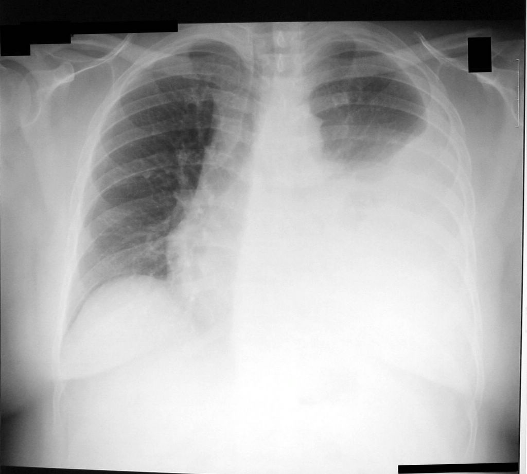
Picture 1 – Pleural effusion
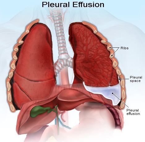
Picture 2 – Pleural effusion Image
If you, or any of your family members, are suffering from symptoms similar to that of Pleural effusion, do not delay treatment. Delay in diagnosis and medical treatment can give rise to a range of complications and jeopardize health. Seeking medical attention on an immediate basis can ensure faster cure of the disease and a quicker recovery.
References:
http://www.ncbi.nlm.nih.gov/pubmedhealth/PMH0001150/
http://www.webmd.com/lung/pleural-effusion-symptoms-causes-treatments
http://emedicine.medscape.com/article/299959-overview
http://www.medicinenet.com/pleural_effusion/article.htm#what_is_pleural_effusion