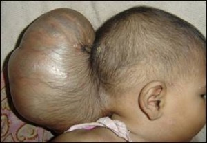
Encephalocele is a life-threatening birth defect that is a subject of many medical researches today. Read and find out all about the disorder, including its causes, symptoms, treatment and prognosis.
Encephalocele Definition
Page Contents
- 1 Encephalocele Definition
- 2 Frontal Encephalocele
- 3 Occipital Encephalocele
- 4 Encephalocele Causes
- 5 Encephalocele Incidence
- 6 Encephalocele Classification
- 7 Encephalocele Symptoms
- 8 Encephalocele Location
- 9 Encephalocele Risk Factors
- 10 Encephalocele Diagnosis
- 11 Encephalocele Treatment
- 12 Encephalocele Surgery
- 13 Encephalocele Prognosis
- 14 Encephalocele Recovery
- 15 Encephalocele Life Expectancy
- 16 Encephalocele Prevention
- 17 Encephalocele Pictures
This is a rare form of neural tube defect (NTD) that affects the brain and is evident at the time of birth. The condition is pronounced as En-Sef-A-Lo-Seal.
The condition is known by various names, such as:
- Cephalocele
- Cranium bifidum
- Craniocele
- Bifid cranium
It is sometimes referred to by the Latin name “Cranium Bifidum”.
Frontal Encephalocele
It is a type of the condition manifested through the front part of the skull. In this type, an abnormal lump protrudes through the frontal skull region. It is more common in people of Southeast Asia. This is a rare type of the disorder.
Occipital Encephalocele
It is a congenital (present at birth) neurological form. It is marked by rare lesions, which are huge in size. These are characterized by progressive inflammation of abnormal masses that protrude through the occipital region of the skull. In many cases, the swelling is present since the time of birth. These lumps can vary from a small polyp-like growth to a huge inflamed mass.
In case of giant forms of this type, the size of the inflamed growth is more than the size of the head that they arise from. Due to the enormous size of these growths, these pose a challenge to surgeons.
Encephalocele Causes
The exact cause of the condition is unknown as yet. However, the disease is found to arise when a meningeal sac comprising of brain tissue projects from the skull. The primary cause for the disorder is an inability of the neural tube to shut down completely at the time of development of the fetus during pregnancy. This results in an aperture in the midplane of the upper skull area, the back of the skull or the section between the nose and the forehead.
In a normal infant, the spinal cord and the brain (central nervous system) develop inside a structure known as the neural tube. A section of the brain may project outside the neural tube if the latter fails to close properly. A sac, either the skin or a thin layer of membrane, covers the brain section jutting outside the skull.
As per medical research, certain substances are found to raise the possibility of this disease. These involve:
- Trypan blue, a stain that colors cells or dead tissues in a blue hue
- Teratogens, which lead to birth defects
- Arsenic, which may cause damage to a developing fetus
Encephalocele Incidence
This is a rare disease and is seen to affect one out of 5,000 live births around the world. The form of this condition that affects the back of the head is more frequent in North America and Europe. The type affecting the frontal head area is more common in Africa, Southeast Asia, Russia and Malaysia. While boys are more susceptible to having the form of the condition affecting the frontal head region, girls stand at a higher risk of suffering from the type that affects the back of their skull.
Encephalocele Classification
The disease is generally categorized into three types:
- Nasofrontal
- Nasoethmoidal
- Naso-orbital
However, the forms of this condition tend to overlap.
Encephalocele Symptoms
The condition is marked by sac-shaped projections of the brain as well as the membranes covering it through the skull openings. Simply put, the brain of an unborn infant may protrude through an opening of the skull. In some cases, section of the membrane covering the brain as well as spinal cord (meninges) and CSF (Cerebrospinal fluid) also come out through the skull aperture. The disorder is frequently accompanied by brain malformations and craniofacial abnormalities.
Some of the main signs and symptoms of this disease may involve:
- Hydrocephalus (High accumulation of cerebrospinal fluid in the human brain)
- Spastic quadriplegia (Paralysis of the limbs)
- Microcephaly (unusually small size of head)
- Ataxia (unorganized movement of the voluntary muscles, like those associated with walking and reaching)
- Seizures
- Developmental delay
- Vision problems
- Retarded mental and physical growth
The severity of the disorder tends to vary on the basis of the region that it affects.
Encephalocele Location
An Encephalocele may be situated in:
- Base of the skull
- Forehead, nose and sinuses
- At any region in the midline around the skull, from the top to its back
Encephalocele Risk Factors
The possibility of having this disorder is likely to be determined by heredity, environmental factors, parental age as well as ethnicity. Individuals having a family history of the disorders spina bifida and ancephaly are also supposed to be more susceptible to this condition.
Encephalocele Diagnosis
The condition is usually detected by doctors during a physical examination of an infant at the time of birth. In some cases, a small Encephalocele may not be evident right after delivery. In such cases, the location is usually in the sinuses, nose or forehead.
In cases where one of the parents-to-be have a family history of the disorder, or are risk-prone in some way, MRI scans, ultrasounds and amniocentesis may be used. These may help detect any possible birth defects or abnormalities. MRI scans can help get accurate images of any congenital defects and associated anomalies.
Encephalocele Treatment
In most cases, the condition is treated through surgical means. Surgery aims at putting the brain section outside the skull back into its original position, and closing the aperture (opening). In some cases, neurosurgeons even manage to repair large lumps of this type without further hampering the functioning ability of an affected child. Reparative surgery, usually performed during infancy, is presently the only effective treatment for this disease.
Treatment usually involves closing open skin defects to avoid drying up of brain tissue and infections. It also aims to remove non-functioning cerebral tissue (outside the cranium) and close the dura tightly. The goal of cure also involves total reconstruction of the craniofacial region. Particular care is given on preventing long-nose deformity.
The success of surgical treatment depends on the size and location of these abnormal masses. In many cases, surgery helps reposition the protruded section of the brain back into the skull. Operative techniques also help remove the projections, repair the deformities and ease pressure on the brain that can hamper normal development of the brain.
In some cases, shunts are positioned to drain away excessive CSF from the brain.
Encephalocele Surgery
Neurosurgeons usually consider a surgical repairing of the disorder within the initial few months after the delivery of an affected infant. If there is a skin covering for the lump, which provides it with some protection, surgeons may prefer postponing the operation for a few months. In the absence of such a covering, an immediate surgery may be recommended. This is likely to be conducted right after birth.
During treatment of this disorder, neurosurgeons generally use an operative procedure known as Craniotomy. The operation involves making an opening through the skull. This surgical technique helps get access to the brain of a child having this condition. During the surgical process, a part of the skull bone is cut off and removed. Following this, surgeons cut the dura mater or the touch membranous covering that protects the brain. After this, the surgeon replaces the brain tissue and any fluids or membranes that project out of the opening of the skull. The sac surrounding this is removed and the dura mater is closed. The aperture of the skull is closed, possibly with the same bone piece that had been removed. In case of a large hole in the surface of the skull, an artificial plate may be used to cover and repair the region.
Encephalocele Prognosis
The outcome of this disorder tends to vary, depending on
- The form of brain tissue that has been affected
- The position of the sacs
- Accompanying malformations of the brain
Babies having a frontal abnormality, no associated defects or syndrome and no herniation of brain tissue into the sac have a decent chance of survival. Around 75% of surviving infants are found to live with varying degrees of mental retardation.
Encephalocele Recovery
The possibility and rate of recovery is difficult to predict before performing a surgery. Recovery actually depends on the success of operation, location of the lumps and the types of brain tissues involved. In cases where developmental and neurological problems occur, specialists aim at minimizing both physical and mental disabilities.
Once the operation has been conducted, an infant can be discharged once it has recovered from surgical effects and is able to have enough food to have a normal body weight. The respiration of the baby should also be checked to ascertain normalcy before it can be brought home. In cases where respiratory equipments are necessary for further management, parents are trained about how to handle and operate the apparatuses and perform emergency measures. Once they are sent home, babies often need follow-up care from various specialists. If parent do not wish to use aggressive therapies, infants can go home with hospital support service.
Encephalocele Life Expectancy
Encephalocele sufferers are often found to have a reduced lifespan. This might be due to factors like:
- Premature birth
- Low birth weight
- Presence of multiple birth defects
- Being of Black or African American descent
The survival rate of sufferers of this disorder tends to differ depending on the location of the defect. Those with a frontal anomaly show a 100 percent survival rate while those with a back abnormality have a 55 percent chance of living.
Encephalocele Prevention
Presently, there is no way known to avoid this condition. However, measures can be taken to reduce the risk of occurrence of this condition. According to some recent studies, adding Folic acid (a B vitamin) to the diet of women who can possibly get pregnant can lower the risk of this condition to a great extent. Women of childbearing age should have a daily intake of 400 micrograms of this vitamin. A single serving of most fortified cereals and multivitamins on a daily basis can supply the body with 400 micrograms of this component.
Doctors also recommend certain general measures, such as abstaining from smoking and drinking during pregnancy, to reduce the risk of their kids from developing this condition.
Encephalocele Pictures
Check these carefully selected images to get an idea about the physical appearance of individuals suffering from this condition.
Picture 1 – Encephalocele
Picture 2 – Encephalocele Image
Fuetuses with this abnormality are likely to die even before birth. Around 21%, or 1 out of 5 fetuses, are said to survive. Only half of these are found to live. If you have given birth to an Encephalocele infant, consult your doctor about the possible treatment options and the chances of survival. Once a diagnosis has been accurately conducted, you are likely to be thoroughly counseled about the possible outcome of the condition. If you wish to continue the pregnancy despite being informed about a poor prognosis, palliative care may be offered to your baby once it is born and in case it manages to survive.
References:
http://lisette-samisblog.blogspot.in/2009/06/what-is-encephalocele.html
http://en.wikipedia.org/wiki/Encephalocele
http://www.ninds.nih.gov/disorders/encephaloceles/encephaloceles.htm


