Epidermolytic Hyperkeratosis Definition
Page Contents
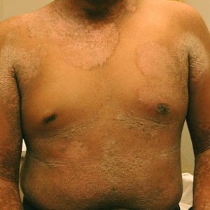
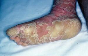
Epidermolytic hyperkeratosis (EHK) is a rare skin disorder arising at birth. It involves the clumping of keratin filaments. It is characterized by generalized erythroderma and severe hyperheratosis with small wart-like scaly formations over the entire body, especially in the body folds and sometimes on the palms and soles.
There is also the presence of recurrent bullae on the lower limbs. Thus, the condition is also known as bullous congenital ichthyosiform erythroderma.
Epidermolytic hyperkeratosis Types
EHK can be divided into two types. People with PS-type epidermolytic hyperkeratosis have thick skin on the palms of their hands and soles of their feet in addition to other areas of the body. People with the other type of epidermolytic hyperkeratosis do not have extensive palmoplantar hyperheratosis. However, they have acquired hyperkeratosis on other parts of the body.
Epidermolytic hyperkeratosis Causes
Mutations or changes in the KRT1 and KRT10 genes are responsible for epidermolytic hyperkeratosis. These genes provide instructions for developing proteins known as keratin 1 and keratin 10. Alterations in these genes result in changes in the proteins of the keratin and hinder them from creating stable and strong network of filaments within the cells. Without a proper network, keratinocytes become fragile and get damaged. This leads to blistering of the skin in response to friction.
Picture 1 – Epidermolytic Hyperkeratosis
EHK is usually inherited in case of an autosomal dominant pattern. This means only one copy of the altered KRT1 or KRT10 gene in each cell is sufficient to cause the disorder.
Epidermolytic hyperkeratosis Symptoms
Babies affected with EHK exhibit very red skin as also serious blisters. Newborns with this disorder usually do not have the protection which a normal skin provides. Thus, they become dehydrated and develop infections in the skin. They may also develop infections throughout the body known as sepsis. The skin is stressed right from birth. It tends to get better after a few weeks.
The scaling starts some months later. Scales are found to develop more in areas that bend or flex. Some patients may also have frequent superimposed infections. The skin gets thickened with age and turns darker than normal. Sometimes, there can be bacterial growth in the thick skin which can give rise to a typical odor.
Epidermolytic hyperkeratosis Diagnosis
The diagnosis of EHK involves:
Laboratory evaluation
Apart from a chosen therapy or bacterial culture, no general laboratory studies need to be done for suspected infection. Keratin defect studies are performed on buccal swabs or blood. If a mutation is identified in an affected individual, mutation-specific testing for relatives and prenatal diagnosis can also be done.
Other Procedures
Skin biopsy is very helpful with histological findings which confirm a diagnosis. Prenatal diagnosis can be made through chorionic villus sampling analysis of amniotic cells or fetal skin biopsies.
Histological Findings
The findings usually observed include thick granular layer, coarse keratohyaline granules and vacuolar degeneration of the upper dermis. Sometimes, the deeper granular cells get dense, enlarged and take shape of irregular masses which appear to be keratohyaline granules.
Patients whose pathologic slides show continuous involvement of the entire horizontal epidermis with these distinctive findings are more likely to have generalized disease. Those who have focal involvement showing skip areas of normal epidermis will possibly have a sporadic, mosaic form of epidermolytic hyperkeratosis. Under electron microscopy, large, round to oval and dense clumps of keratin tonofilaments can be observed in the lower epidermal layers.
Epidermolytic hyperkeratosis Treatment
The skin of an EHK sufferer should be moisturized properly, which is the first step to manage symptoms. Topical emollients should be applied on a regular basis in order to prevent scaling. Taking long and frequent baths is another recommendation for proper skin care. Sea salt can be added to bathwater to have a softening effect. It also makes it easier to do away with the excess skin related to the thickening caused by the disorder. Patients can also use anti-bacterial soap or chlorhexidine which contains cleansers.
Picture 2 – Epidermolytic Hyperkeratosis Image
If the skin gets irritated or develops associated blisters become inflamed, the doctor might prescribe oral medications. Antibiotics are given to control any infection which might be present. Anti-inflammatory medications are used to control any swelling or inflammation of the affected skin. Oral retinoids such as isoretinoin are used to manage the symptoms of epidermolytic hyperkeratosis. Isotretinoin is derived from Vitamin A and is known to be the miracle drug.
Epidermolytic Hyperkeratosis Prognosis and Follow-Up
Once treatment is over, follow-up visits to doctor are required for symptomatic relief. Follow-up laboratory studies might be needed during systemic therapies for EHK. For treatment of neonatal patients, an affected baby may be taken to ICU to be watched for sepsis and electrolyte imbalance when required. EHK patients must be properly cared after as they have a risk of developing infections again.
References:
http://emedicine.medscape.com/article/1112403-overview
http://ghr.nlm.nih.gov/condition/epidermolytic-hyperkeratosis
http://en.wikipedia.org/wiki/Epidermolytic_hyperkeratosis
http://medical-dictionary.thefreedictionary.com/epidermolytic+hyperkeratosis


