What is Tracheal Deviation?
Page Contents
Tracheal Deviation: The trachea, more commonly known as windpipe is one of the most important parts of the body as it is used for breathing. Its structure is like a tube which is about 1 inch in diameter and 4 to 6 inches in length. It is vertically located in front of the esophagus having 16-20 cartilage rings that prevent it from collapsing.
The trachea is a membrane suppurated by cartilage rings having an opening at the back. It is connected by a band of muscles which in return helps the trachea to expand and contract whenever we breathe in and out. It also acts as a defense mechanism whenever it is exposed to various environment irritants. While coughing, the mucous membrane that is lined on the inside becomes very sensitive, stimulates reflex action and coughs up the foreign body which enters the body.
Causes of Tracheal Deviation
- In a relatively high negative pressure, the tracheal shifts on either of the sides resulting in tracheal deviation.
- Situated in a vertical position in a straight manner starting from the neck area to the chest area, a deviation in its position will highly affect the area
- Pneumothorax as it is said to be the most dangerous condition that causes it
Tracheal Deviation in Infants
Tracheal deviation in infants is not a major issue and has no medical concern. Infants up to the age of five years are prone to have a tracheal deviation that has no link to medical conditions. It happens because the infants have short neck relatively to trachea which resolves itself and outgrows afterward.
- Atelectasis: Collapsed lung in medical terms is considered as atelectasis that happens due to an obstruction or blockage that restricts its capacity to inflate adequately.
- Pleural Fibrosis: The pleura (the lining of the chest cavity) gets so thickened that it results in chronic inflammation to calcification.
- Pleural Effusion: It is a condition when fluid gets trapped in the chest wall and lungs. Although tracheal deviation is a prominent researched case, its symptoms can only be seen once there is a huge amount of fluid collected.
- Tumors: Either of the benign or malignant can cause tracheal deviation. A large mass found in the lung, bronchi or pleural cavity can displace the trachea to the side.
- Pneumothorax: It may arise when there is an injury to the lungs. Due to the large quantity of air entering the pleural space, the lung may collapse. During respiration, this air gets compressed and restricts the expansion of the lungs which in turn leads to other life threatening problems.
Signs and Symptoms
Respiratory symptoms
- Respiratory distress or respiratory arrest also related to cardinal finding
- Increase in the rate of respiration
- Lung expansion Asymmetry
- Decrease in breathing sound or complete absence of it
- Breathing sounds Wheezes or crackles (also called adventitious breath sounds)
- Difficulty in breathing (dyspnea)
Cardiac Findings
- Tachycardia increase in cardiac rate
- Hypotension decrease in blood pressure
- Dented Jugular vein
Other Findings
Detection of Tracheal Deviation & other Signs
Grossly examining or a mere palpation will easily detect the deviation of tracheal. Even a mere x-ray report will reveal the following:
- Dyspnea (difficulty in breathing)
- Cough
- Abnormal breath sounds
Treatments for Tracheal Deviation
Tracheal deviation by itself does not cause any major health issue. But its causes and symptoms are the underlying problems that need to be addressed. Moreover, it is always treated with many precautions to avoid risks that may even lead to death.
- Needle Thoracostomy: 14-16 cannulas that are inserted on the second intercostals space which is advanced until the air can be released through the syringe connected to the cannula. As soon as it is set in place, a guiding needle will be removed to let the air trapped inside to escape.
- Chest Drain Placement: It is one of the most popular and common treatments used for pneumothorax and are surgically done. A tube is placed on the pleura that allow the air to escape. This process is considered to be safer since this is done visually compared to the needle thoracostomy
Tracheal Deviation Pictures
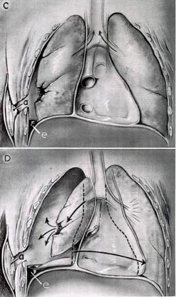
Tracheal Deviation image