Albinism generally encompasses a group of hereditary conditions that involve unusual cases of hypopigmentation. Get to know all about this condition in detail, including its causes, symptoms, diagnosis, treatment and prognosis.
Albinism Definition
Page Contents
It is a congenital disorder marked by partial or total loss of melanin, a pigment naturally found in the hair, skin and eyes. In this condition, the melanocytes responsible for producing melanin are completely damaged or absent. This is completely reverse or opposite of melanism, in which the skin undergoes dark pigmentation.
The disorder is also known by several other names like:
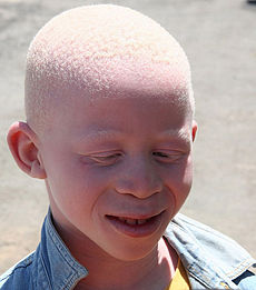
Picture 1 – Albinism
- Achromatosis
- Achromia
- Achromasia
Albinism Incidence
The frequency of the disorder is approximately 1 in 17,000 to 1 in 20,000. Although the condition affects all ethnic populations, most cases are reported from the tropical regions, particularly Africa. It is estimated that over 150,000 achromia-affected individuals live in Tanzania out of which 8,000 are registered with the Tanzania Albino Society.
Albinism generally has a huge psychological impact on the sufferers due to the non-cooperative attitude of the society. It is highly essential to educate the general public as well as the family members of patients on this condition. The various facts and myths related to the condition should be known to everyone. This is extremely important as patients require extensive support and help from the society in order to lead a normal social life. A number of organizations like NOAH and fellowships are available for patients to cope with the problem. As the condition follows an inheritance pattern, prenatal screening and genetic testing are increasingly used by the carriers of the mutant gene.
Albinism Types
The condition may exist in either of the following two forms:
Oculocutaneous Albinism
In this type, pigmentation of the skin, eyes, and sometimes the hair gets severely affected. The condition could be further classified into the following types:
- OCA 1
- OCA 2
- OCA 3
- OCA 4
Ocular Albinism
Unlike the previous form, this type of the condition particularly involves the eyes. Patients with this less common disorder exhibit normal skin and hair. It usually manifests into any of the following variants:
- OA 1
- OA 2
- OA with sensorineural deafness
Albinism Symptoms
Impaired synthesis of melanin can lead to a varied range of symptoms in both types. The oculocutaneous disorder is characterized by an abnormal discoloration of the hair. Affected skin appears white or pale in contrast to the normal color. This could be, however, detrimental to the skin due to constant exposure to the ultraviolet radiation (UV rays) from the sun. Therefore, albino patients often complain of skin burns and discomfort after a prolonged or intense sun exposure. In addition to this, few patients may develop abnormalities like:
- Pigmented or non-pigmented raised moles
- Lentigines, which are the tiny, pigmented spots with clearly-defined edges
- Small tan or light brown spots called freckles
Since melanin is also responsible for pigmentation of the iris to increase the opacity of the eye, its deficiency results in a red or purple retina. A translucent iris loses the ability to prevent the light from entering the eye and causes increased photosensitivity, also called photophobia. Failure to produce sufficient amount of pigment can lead to a severe deterioration of the optical system, causing a host of visual defects. Some of these include:
Decreased visual acuity
Affected individuals have a lack of clarity or clearness of vision due to scattering of light within the eye.
Macular hypoplasia
In this condition, improper development of the oval-shaped yellow spot near the retina (called macula) affects the vision. This could be attributed to an underdeveloped pigmented layer of retina called retinal pigment epithelium, which is a common feature in albino patients.
Retinopathy
Excessive exposure of the eyes to the UV rays can damage the retina.
Optic chiasma
Non-pigmentation of the iris often results in crossing of optic nerve fibers.
Amblyopia
Partial or complete loss of vision may occur in one eye due to inadequate stimulation and poor development of brain cells in the visual cortex.
Astigmatism
Irregular shape of the cornea may cause visual blurring and discomfort.
Optic nerve hypoplasia
In this case, the optic nerves are underdeveloped.
Nystagmus
Many patients develop this unusual condition in which the eyes undergo voluntary or involuntary movements, causing reduced vision.
Albinism Causes
Deficit in the production of melanin is a hereditary disorder. The gene responsible for encoding tyrosinase, an enzyme that regulates the production of melanin, undergoes mutation. The inherited disease usually follows a recessive pattern, which means the condition appears only in those individuals who have received two copies of the mutant gene, one from each parent. Although the pattern of inheritance is the same in both types of achromia, the location of the defective gene may change. The several versions of oculocutaneous albinism display the following features:
Type 1
The mutation of the tyrosinase gene occurs in chromosome 11q, resulting in complete absence or reduced activity of the enzyme.
Type 2
In this case, the melanosomal tyrosine transporter (called P polypeptide) is defective. The P gene is usually localized to chromosome 15. Affected patients may have traces of melanin, which gives rise to partial coloration.
Type 3
The tyrosine-related protein-1 gene (TYRP1) on chromosome 9p undergoes mutation. Pigmentation of the iris occurs in the first decade of life, despite the absence of tyrosinase. The condition is sometimes called rufous oculocutaneous albinism.
Type 4
In this form, the anomaly lies in the SLC45A2 protein that assists in the functioning of tyrosinase. Hence, the albinistic individuals often produce slight amount of the pigment.
Type 1 ocular albinism is a typical example of an X-linked recessive disorder in which the OA1 receptor gene known for producing intracellular G-protein coupled receptor-like protein undergoes mutation. The most significant characteristic of the condition is the presence of large-sized melanosomes in the skin. A mutant CACNA1F gene shares a strong relationship with type 1 of the ocular disorder.
Albinism in animals
This pigmentation disorder also affects a large number of animals like:
- Humpback whales
- Bottlenose dolphins
- Gorillas
- Penguins
- Buffalos
- Elephants
- Deers
- Alligators
- Rabbits
Albinism Diagnosis
Physicians always go for a thorough assessment of the skin, hair and eyes of patients to evaluate the underlying cause. The pattern of pigmentation needs a careful analysis, owing to the existence of multiple variants. A few diagnostic approaches need to be followed for confirming the congenital disorder.
Genetic testing
In this procedure, the several genes involved in the production of melanin are sequenced one by one to detect the type of the disorder.
Hair bulb incubation test
It is an important test that helps in distinguishing a tyrosinase-positive patient from a tyrosinase-negative one. The method involves extracting hair shafts with follicles from the scalp of a patient. The follicles are then incubated in a test tube containing a solution of L-dopa or L-Tyrosine that facilitate melanin production. If the hair follicles turn brown or black, the condition is marked tyrosinase positive.
Skin/Hair bulb biopsy
A histological examination of the hair follicles or skin may aid in finding out the presence of melanosomes as in the case of Type 1 ocular albinism.
Neuroimaging studies
The visual-evoked potential (VEP) generated by the optical nerves can be recorded by examiners to detect the condition. Individuals affected by Albinism show a delay in the recording of VEP due to disturbance in the transmission of signals through the optic nerve. Optical coherence tomography may help in identifying defects in the retinal structures by differentiating the layers of retina, and determining its overall thickness.
Ocular biopsy
Microscopic examination of the ocular tissues may help in finding out melanin-deficient retina as well as presence of lipid deposits in the iris.
Albinism Differential Diagnosis
During the diagnosis, it is mandatory for examiners to ensure the absence conditions that give rise to similar symptoms. Some of these include:

Picture 2 – Albinism Image
- Hermansky-Pudlak Syndrome
- Chediak-Higashi Syndrome
- Aniridia
- Congenital or infantile esotropia
- Prader–Willi Syndrome
- Angelman’s Syndrome
Albinism Treatment
Visual rehabilitation could help in correcting the ocular defects up to a certain extent. Several methods are specifically designed to enable patients to make an optimum use of their low vision. Although albinistic individuals undergo a limited spontaneous recovery within the first few months, the existing visual anomaly may remain as a permanent problem. Ocular surgery may help in the proper alignment of the eyes in patients with a squint. The visual errors associated with nystagmus and astigmatism may require prompt surgical repair. Bifocal lenses provide an optimal balance between distance vision and near vision depending on the severity of the visual defect.
Health specialists may recommend patients, especially young children to use prescription reading glasses, magnifiers or monoculars. Colored contact lens aid in alleviating photophobia, but should not be worn by individuals with nystagmus. A pair of vision-enhancement lens called bioptic telescope is a reliable optical aid that improves distance vision in patients with ocular disability. Patients who are having increased light and skin sensitivity must take few precautionary measures like:
- Wearing sunglasses or tinted visual aids before going out in the sun
- Applying sunscreens on solar damage-prone areas prior to an outdoor activity
- Wearing protective clothing and brimmed hats to prevent overexposure to the UV rays
Albinism Prognosis
The disorder is absolutely not associated with any fatal outcome. Most albinos have a normal growth and development. However, lack of synthesis of melanin can allow the UV rays to penetrate deep into the skin, increasing the risk of cancer.
References:
http://en.wikipedia.org/wiki/Albinism
http://www.albinism.org/publications/what_is_albinism.html
http://emedicine.medscape.com/article/1216066-overview
http://www.ncbi.nlm.nih.gov/pubmedhealth/PMH0002450/